

Journal of Cell Biology
@JCellBiol
Followers
50K
Following
5K
Media
10K
Statuses
15K
The Journal of Cell Biology (JCB) publishes peer-reviewed research on all aspects of cellular structure and function. Est. 1955 - @RockUPress
New York, NY
Joined September 2009
Our annual collection highlights twelve of the most outstanding papers that showcase the exceptional range and quality of research published by JCB. We are delighted to serve as an independent venue for the awe-inspiring world of cell biology! #CellBio2024

1
5
22
.@samjlord @KatrinaVelle @MullinsLab & @FritzLaylin propose a simple way to highlight both experimental #reproducibility and cell-to-cell variation, while avoiding pitfalls common in analysis of cell biology data.

4
170
336
.@kat_shkarina, @broz_lab @unil and colleagues report the development and characterization of a new optogenetic toolset for #apoptosis, #necroptosis, and #pyroptosis induction in humans, mice, and #zebrafish. #CellDeath #optogenetics #Immunology
2
76
286
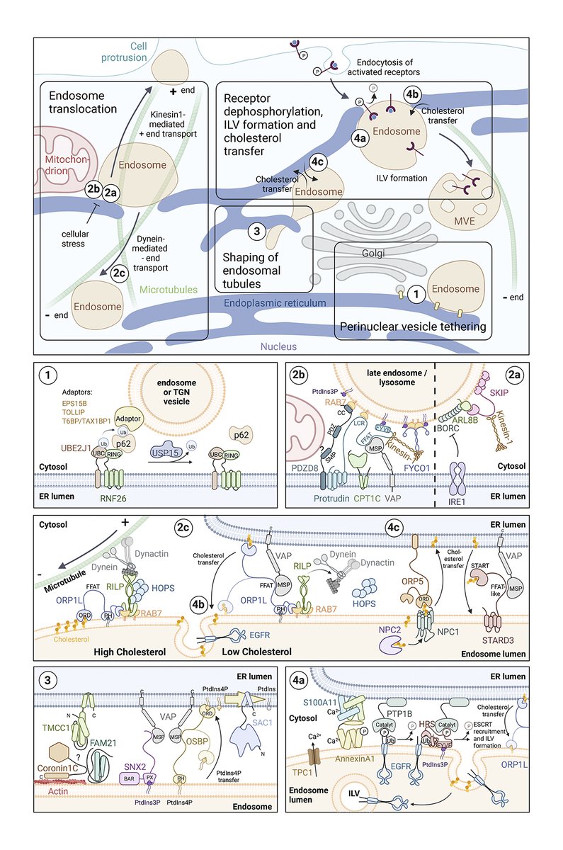
.@EvaMariaWenzel, @harald_stenmark et al. review how the endoplasmic reticulum controls the positioning, dynamics, & functions of other #organelles via membrane contact sites. Part of our #Lipid and #Membrane Biology collection:

0
98
291
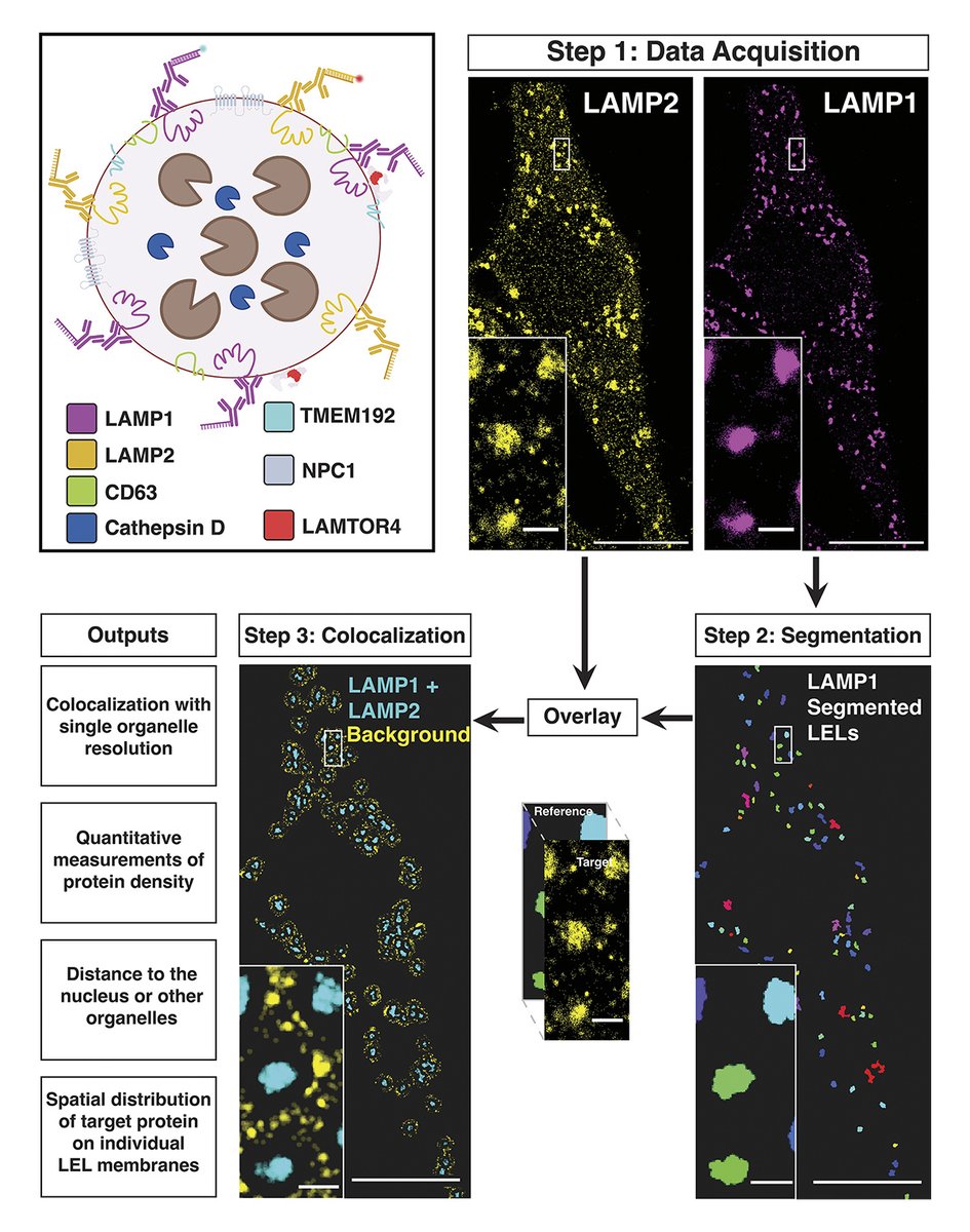
New work from @Charlie_R_Bond & @Melike_Lak et al offers multiplexed DNA-PAINT super-resolution pipeline for investigating #organelle heterogeneity with single organelle resolution, identifying up to 8 subsets of late #endosomes/#lysosomes in a single cell
2
52
245
Takami Sho, Li Yu et al. @Tsinghua_Uni unveil migratory autolysosome disposal, a response to #lysosome damage where cells expel LAMP1-LC3 positive structures via autolysosome exocytosis, requiring #autophagy machinery, SNARE proteins, and cell #migration.
0
59
222
This poster-as-DM-thread from Jimmy Hsu in @GaryBrouhard ’s lab has got to be the most innovative poster idea I’ve seen! And it’s not just a gimmick - it clearly communicates some really cool science about microtubule dynamics. (Photo poster with permission.) #mweJCB #ASCBEMBO19
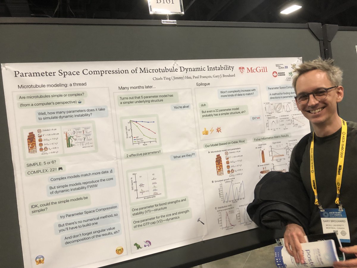
4
51
186
Wenzel, Elfmark, Stenmark, and Raiborg @Oslounivsykehus review how the endoplasmic reticulum controls the positioning, dynamics, and functions of other #organelles via membrane contact sites. #ER_literature #Biochemistry #Trafficking #Metabolism

0
55
190
We are pleased to introduce the members of the 2024 JCB Early Career Advisory Board. This initiative serves the journal’s ongoing goal to be representative of and responsive to every part of the cell biology community.

2
20
158
.@XiaoLinichd, Gamuyao, @chiluncellbio et al. @stjuderesearch engineer a #LipidDroplet (LD)-targeting motif and develop a tool kit that can track contact sites between LDs and other organelles in living cells. @Daniel_Stabley @alexcarisey @agtebo

3
45
160
Using live imaging, optogenetics, & laser-induced tight junction injury, @saranya_raajan, Ann Miller et al @UMich_MCDB show that mechanosensitive channel-dependent calcium flashes promote sustained local activation of RhoA and tight junction repair
0
41
150
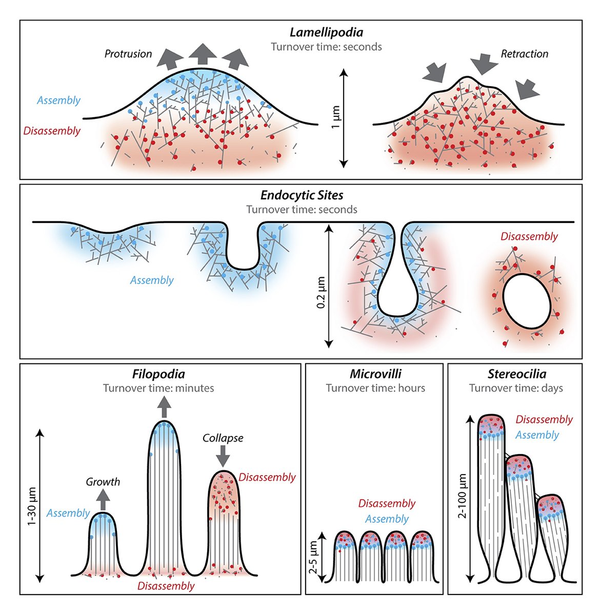
.@BruceLaneGoode, Eskin and @sshekhr review our current understanding of the cellular machinery and mechanisms driving actin network remodeling, disassembly, and turnover. #Cytoskeleton #Biochemistry
1
42
148
JCB is pleased to present a special collection highlighting recent papers on the mechanical properties of cells and tissues, and the application & transmission of forces across scales relevant to cell biology. Gain new insights into #mechanobiology 👉

0
43
141
Image shows accumulation of mtDNAs in the cytosol stimulates the cGAS-STING-mediated immune response. In this review, Sookyung Kim, Theresa Ramalho, and @chaynes329 discuss how #mitochondria-to-nuclear communication promotes #proteostasis and #immunity.

0
37
144
Tight junctions regulate the mechanical resistance of epithelial cell junctions. @tann_nguyen, @tetsuotani1, Mikio Furuse et al. show that cell junctions fracture upon cell stretching in cells lacking TJ membrane proteins claudin, JAM-A, and CAR.
1
31
137
#Optogenetic activators of #apoptosis, #necroptosis, and #pyroptosis. Kateryna Shkarina (@kat_shkarina), @broz_lab @unil and colleagues report the development and characterization of a new optogenetic toolset. #Optogenetics #CellDeath #Immunology
0
26
134
Endogenous gene tagging by #PCR in mammalian cells made easy. #CRISPR #Cas12 enabled #GeneEditing with a comprehensive toolbox on @Addgene. Collaboration by the @LabKnop, @lemberg_lab and @PereiraLab1 @ZmbhH and @DKFZ

1
49
136
Ever wonder about the mechanics of immune cell #migration? @AlexiaCaillier, @pwoakes @LoyolaChicago and colleagues find T cells form integrin mediated focal adhesions to pull themselves through confined spaces. #Immunology #Adhesion

0
42
140
Video shows tomogram of an endosome in vps13Δ cells. Sho Suzuki (@ShoSuzuki20) @CornellMBG et al propose that Vps13 may play a critical role in supplying #lipids to #endosomes, ensuring continuous ESCRT-mediated sorting during MVB biogenesis. #Trafficking
0
22
136
Fat is stiffer and more dangerous than you think, especially to the nucleus of a cell. Ivanovska and Tobin in the Discher lab @BB_UPenn measure the physical effects of fat droplets within cells. #Membrane #lipid #Biophysics #LipidDroplets

0
40
132
A live-cell marker to visualize the dynamics of stable #microtubules throughout the cell cycle. From Klara Jansen, Malina Iwanski @mkiwanski, Mithila Burute and Lukas Kapitein @KapiteinLukas @UniUtrecht: #Technology #Cytoskeleton
0
31
133
.@kat_shkarina, @broz_lab et al report the development & characterization of an #optogenetics toolset to induce #apoptosis, #necroptosis & #pyroptosis in humans, mice & #zebrafish From the Year in Cell Biology 2022: #CellBio2022
1
24
129
ER as master regulator of membrane trafficking and organelle function. A new review from Eva Maria Wenzel, Liv Anker Elfmark, @harald_stenmark, and Camilla Raiborg @Oslounivsykehus: #Biochemistry #Organelles #Trafficking #Metabolism #ER_literature
2
29
135
To assist researchers and institutions adjusting to working remotely, we are making all articles in @JCellBiol free to read until May 31, 2020. Learn more about the steps we’re taking and find COVID-19 relevant content:
1
63
122
New #optogenetic tools developed by @kapiteinlukas and colleagues @UniUtrecht enable reversible repositioning of cellular #organelles to dissect intracellular transport and spatial cell biology
0
32
117
The E4 variant of APOE is the strongest genetic risk factor for late-onset #Alzheimers. @ia_windham et al @cohenlaboratory find that APOE can traffic to the surface of #LipidDroplets in astrocytes. APOE4 on LDs causes defects in LD composition & morphology

0
43
124
Orii and Tanimoto @YCU_koho study the structural mechanics of #microtubule and #actin cytoskeletons using high-force intracellular magnetic tweezers. The measurements directly reveal the integrated nature of the two cytoskeletons throughout the cytoplasm
2
32
126
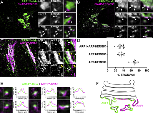
.@freethepipette, @fran_bottanelli and colleagues combine gene editing and STED super-resolution #microscopy to reveal the nanoscale localization of ARF #GTPases in living cells
1
32
122
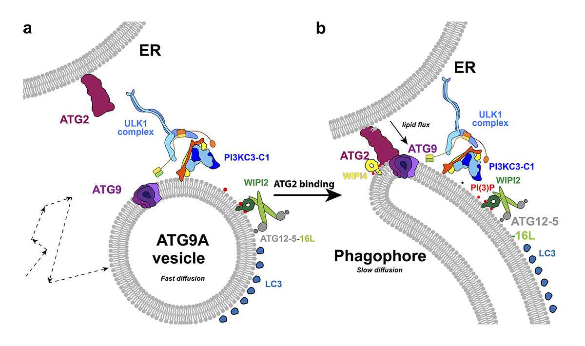
Spotlight: Cook and @TheHurleyLab @berkeleyMCB preview two related studies (@tjolivas et al. & @DavidGBroadbent et al. , examining the role of ATG9A in autophagosome biogenesis in mammalian cells #autophagy
1
30
122
This month, browse a collection of classic papers on #mitochondria for #JCB65. The collection encompasses many groundbreaking studies examining mitochondrial structure, function, and dynamics

3
56
117
Using a combination of modern cell biology techniques, including #trafficking assays, #CRISPR, and secretomics, Pereira, Stalder, @dgershlick et al. @Cambridge_Uni reveal that the exocyst complex has a fundamental role in protein secretion.
2
30
111
How does mitochondrial dysfunction in one cell impact neighboring cells? This study by Yipeng Du, Matthew Sieber et al. @UTSWMedCenter shows that mitochondrial ROS production regulates membrane receptor trafficking and disrupts cell-cell signaling.
1
28
120
.@AmeeeenNeuro @guiwitz @ben808080 et al. @unibern developed CryoVesNet, which enables accurate segmentation of synaptic vesicles in cryo-electron tomograms. This tool advances our understanding of synaptic function and synaptic vesicle pool dynamics.
1
27
113
APOE targets cytoplasmic #LipidDroplets via the ER and modulates LD composition and size. @ia_windham et al. @cohenlaboratory @UNC find that APOE can traffic to the surface of lipid droplets in #astrocytes. #Neuroscience #lipid #Metabolism #Alzheimers

0
24
115
.@TobiasKletter, @SimoneReber et al. introduce Spindle3D, an open-source image analysis tool that allows for consistent and automated morphometric quantification of the mitotic #spindle and #chromatin Part of our Tools collection:

1
24
116
Klara Jansen, @mkiwanski, @KapiteinLukas et al. @UniUtrecht introduce StableMARK (Stable Microtubule Associated Rigor-Kinesin), a live-cell marker to visualize stable #microtubules. #Technology #Cytoskeleton
0
22
113
A new probe for phosphorylated RNA polymerase II enables visualization of the sites of transcription in living cells. From Satoshi Uchino, Yuma Ito, Hiroshi Kimura @tokyotech_en and colleagues: #Technology #Biophysics #Chromatin #epigenetics
0
31
109
Heterogeneity of late #endosome/#lysosomes shown by multiplexed DNA-PAINT imaging. New quantitative pipeline developed by Charles Bond (@Charlie_R_Bond), Melike Lakadamyali (@Melike_Lak) and colleagues @PennMedicine: #Biophysics #Organelles #Technology

3
26
113
Monster et al. @GloerichLab show how epithelial cells can round up in #mitosis while maintaining epithelial barrier integrity. This requires an asymmetric composition of mitotic cell-cell junctions through an E-cadherin mechanoresponse in neighbor cells.
2
30
104
Ani Michaud, @GoryachevAndrew, George von Dassow, William Bement @BementLab, et al. identify a versatile cortical pattern-forming circuit based on #Rho, F-#actin, Ect2, and RGA-3/4. #CellCycle #CellDivision #Cytokinesis #cytoskeleton
0
27
98
Fujiwara, Kusumi et al. developed an ultrafast camera system that enables the highest time resolutions in single fluorescent-molecule imaging to date: In our Tools collection:
1
30
106
Gaia Pigino (@GaiaPigino) @mpicbg studies the molecular mechanisms and principles of self-organization in #cilia using 3D cryo-electron microscopy. Learn more about her work in our latest #PeopleAndIdeas:

4
17
103
#Microtubule and dynein-mediated removal of cortical myosin II promotes polar relaxation and facilitates assembly of the actomyosin contractile ring #cytokinesis #anaphase @SPOREMOHAN @midbody
1
26
104
.@ShoSuzuki20 et al @CornellMBG reveal a critical role for Vps13-mediated #lipid transfer at ER–endosome contact sites in ESCRT-mediated sorting, elucidating the contribution of the ER to membrane protein sorting at #endosomes #Trafficking #ER_literature

1
28
111
.@ShoSuzuki20 et al. propose that Vps13 may play a critical role in supplying #lipids to #endosomes, ensuring continuous ESCRT-mediated sorting during MVB biogenesis. From our Inter-Organelle Contacts collection:
1
25
108
The heterogeneity of lysosomes within a single cell has been challenging to capture and study in detail. Chen & @maxgabgut discuss new work by @Charlie_R_Bond, @Melike_Lak et al ( that tackles this question using DNA-PAINT imaging:

0
31
110
ER–plasma membrane contact sites deliver ER lipids and proteins for rapid cell surface expansion, say Madison Smith, Lincoln Gay, and Markus Babst @UofUBiology: #Membrane #lipid #Trafficking #Biochemistry #ER_literature
0
24
106
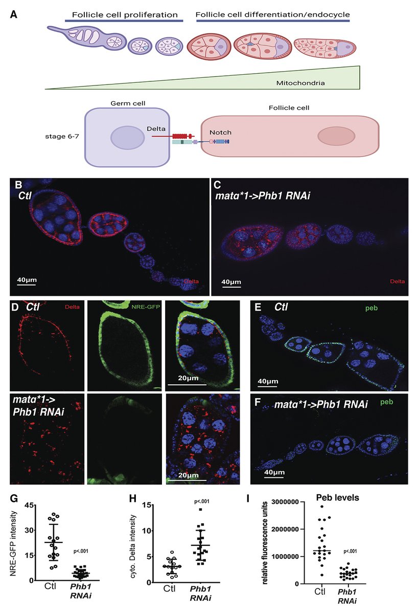
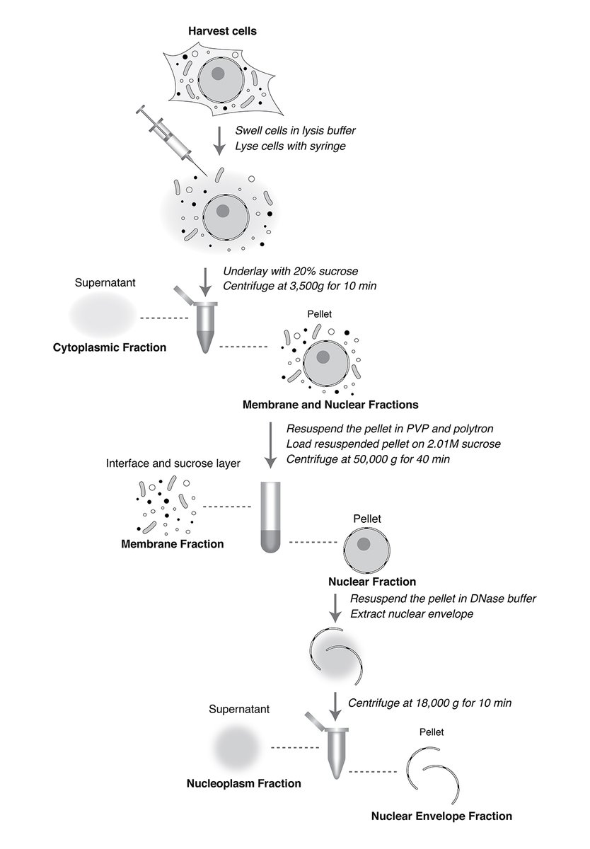
.@udi_yael et al. @RoutLab_RU present a new method for quantitative subcellular fractionation of a wide range of mammalian cells while maintaining nuclear integrity. In our Tools collection:
0
24
105
alpha-catenin regulates #integrin adhesions and ECM mechanosensing. A new study from @aavi1907 @Haguy_Wolfenson and colleagues #Mechanobiology #CellBiology #FocalAdhesion #Adhesion #Biophysics
1
20
104
Spatial analysis reveals the location of late endosome/lysosome subsets with respect to the nucleus, mitochondria, & TGN. @Charlie_R_Bond, @Melike_Lak and colleagues @PennMedicine offer insights into organelle heterogeneity at single-organelle resolution.

0
21
102
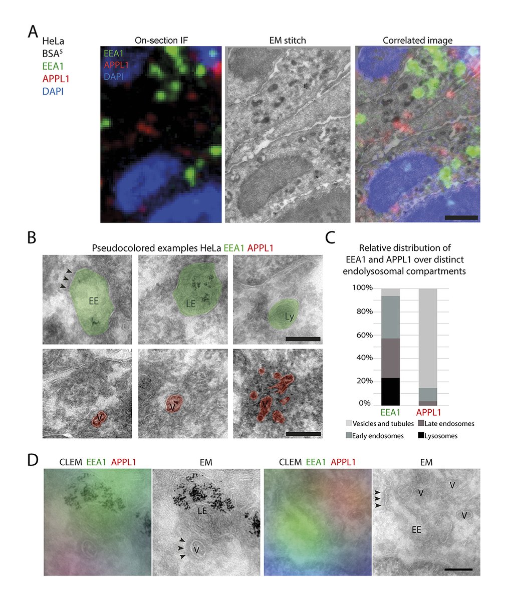
A quantitative CLEM method by @JA_Beek, de Heus, @NalanLiv & @JKlumperman determines the ultrastructural distribution of Rab5, Rab7, EEA1, APPL1 & PI(3)P, revealing distinct endosomal subpopulations & an unexpected localization of EEA1 to late #endosomes
0
27
98
.@LuskingL and @TJ_MeliaLab @yalecellbio suggest an outside-in model of nuclear #autophagy that employs an outer nuclear membrane cargo adaptor, Atg39, that captures inner nuclear membrane into intralumenal vesicles for delivery to the autophagosome.
0
33
100
Inhibition of endolysosome fusion increases #exosome secretion, say Ganesh Vilas Shelke @ganeshs98367397, Chad Williamson @cd_w1, Michal Jarnik, and Juan Bonifacino @BonifacinoJuan @NICHD_NIH: #Disease #Trafficking #Organelles #Cancer

0
22
98
On our January cover: #mitochondria–ER contacts (white) in a COS-7 cell are speckled across the mitochondrial surface (purple) & are easily visualized in the absence of the ER (green). By @BenCardoen et al. JCB’s January issue ➡️
0
23
92
ER-#lysosome #lipid transfer protein VPS13C/PARK23 prevents aberrant mtDNA-dependent STING signaling, say William Hancock-Cerutti, Pietro De Camilli @PDClab and colleagues #Neuroscience #Organelles #Immunology #Membrane #Parkinsons #ER_literature
0
18
100
Feng, Cai, Xu et al. @ZJU_China report that saturated fatty acids cause mitochondrial damage and ROS elevation in human endothelial cells. Activation of #lysosome biogenesis using TRPML1 agonists is sufficient to mitigate SFA-induced #mitochondria damage.

0
25
101
.@tjolivas, Yumei Wu, @shenliang_yu, @TJ_MeliaLab et al. @YaleCellBio show that ATG9 vesicles are the membrane seed for mammalian autophagosomes using nanodisc technology @PDClab.@GuptaLab1.@1in_1uan.@em_guinn.@ShantaNag1. #autophagy

1
28
97
In eukaryotes, VPS13 and related proteins act as channels to transfer #lipids between #organelle membranes, say PeiQi Li, et al. (. From our special collection on #StructuralBiology:

1
20
92
Video shows #optogenetic induction of apoptosis in #zebrafish larvae. @kat_shkarina, @broz_lab @unil et al. demonstrate optogenetic activators of #apoptosis, #necroptosis, and #pyroptosis #Biochemistry #CellDeath #Optogenetics #Immunology
0
14
86
Determining how many cells to average for statistical testing of #microscopy experiments. A Viewpoint from Adam Zweifach @UConnMCB:

0
23
86
.@dombingham et al from @christlet's lab use bead-induced presynapses and CRISPR-tagging of endogenous #actin coupled to STORM to reveal distinct actin nanostructures within presynapses: a mesh at the active zone, rails, and perisynaptic corrals. @Dr_majio

2
33
90
.@alexandraflong @poojasuresh47 & @DumontLab show that individual #kinetochore-#microtubule fibers locally dissipate force to maintain robust mammalian #spindle structure #CellDivision
0
23
90
Rediscover some of JCB’s most-read recent reviews about #mitochondria biology in this new special collection:

0
37
88
Arnold, Riegger, @mixmue et al. @goetheuni present hGRAD, a “one-fits-all” system that can be inserted in one step into any cell that expresses GFP-tagged proteins, allowing their rapid and inducible degradation. #RNAbiology #Biochemistry

0
34
88
.@TobiasKletter, @SimoneReber et al. @IRILifeSciences introduce Spindle3D, an open-source image analysis tool that allows for consistent and automated morphometric quantification of the mitotic #spindle and #chromatin in confocal images.

0
26
83
Cell migration orchestrates migrasome formation by shaping retraction fibers, say Changyuan Fan, Yaming Jiu and colleagues. #Migration #motility #Physiology #Biophysics #Organelles
0
18
88
Mu A, Henry Higgs @hhiggslab @GeiselMed and colleagues review the emerging field of cytoskeletal lysine acetylation, and discuss impacts of #actin acetylation on its cytoplasmic and nuclear functions. #Biochemistry #CellSignaling #Cytoskeleton

0
37
88
Rabas, Palmer, Norman et al. @CRUK_BI show that the metabolic state of #cancer cells can lead to release of #exosomes containing mitochondrial DNA which cause other cells to behave in a more invasive fashion. #CellMetabolism
0
17
93
Quintanilla (@fluoroforce), Oakes (@pwoakes), Beach (@myosincity) and colleagues @LoyolaChicago demonstrate subcellular biophysical mechanisms that enable myosin 2 filament assembly. @MatthewAkamatsu @Coronin @taraskalab .#Biophysics #Cytoskeleton
0
22
87
Nayanika Sengupta, Somnath Dutta @iiscbangalore and colleagues report the high resolution cryo-EM structure of oligomeric VCC, a pore-forming toxin from Vibrio cholerae, in a near-physiological lipid bilayer environment. #Membrane #lipid #StructuralBiology

4
12
92
Jones, Devenport et al. @PrincetonMolBio describe a new mouse model for live visualization of the basement membrane (BM) in which endogenous type IV collagen is fluorescently labeled. #Adhesion #Development

1
22
85
.@dombingham, @christlet et al. use bead-induced presynapses & CRISPR-tagging of endogenous #actin coupled to STORM to reveal distinct actin nanostructures within presynapse. From the Year in Cell Biology: #Cellbio2023

0
14
85
#Mitochondria as intracellular signaling platforms in health and disease - A new review by Jay X. Tan and Toren Finkel @AgingPitt

0
36
87
Integrated model of autophagosome, autolysosome, and lysosome transport along axons. From @sydneycason & @ErikaHolzbaur showing that axonal transport of autophagosomes is regulated by dynein activators JIP3/JIP4 and ARF/RAB GTPases #Neuroscience #autophagy

0
23
90
No microtubules? No problem! @KatrinaVelle and @FritzLaylin show Naegleria use #actin and Arp2/3 complex to crawl and eat, revealing an evolutionarily ancient origin for these phenotypes. #Cytoskeleton #Motility

0
23
84
Integrating recent findings, Zhiming Chen and Sandra L. Schmid @slschmid_lab present a more dynamic, flexible and non-linear model for clathrin-coated vesicle formation.#biochemistry #StructuralBiology

2
27
82
You-An Su, Ya-Wen Liu et al. show that NME3 is a mitochondrial tethering complex that binds to phosphatidic acid derived from cardiolipin, selectively tethering #mitochondria w/ externalized cardiolipin to promote their fusion for functional homeostasis
0
21
87
Video shows that F-actin is necessary for glucose-induced arrest of neuronal mitochondrial motility. @himanishbasu, @GulcinPekkurnaz, Schwarz et al show that FHL2 anchors #mitochondria to #actin & adapts mitochondrial dynamics to glucose supply
1
18
81
Ralhan, Chang, Lippincott-Schwartz @JLS_Lab and Ioannou @IoannouLab review the emerging role of #LipidDroplets in the nervous system in development, aging, and #disease. #Membrane #lipid #Neuroscience

1
24
86
Fuentes and Marin (@zacsimile) et al. @bewersdorflab @YaleCellBio use super-resolution microscopy to reveal that Rtn4 oligomers possess a unique nanoscale organization that helps shape elliptical ER tubules. @jtysonphd @DavidBaddeley5 .#Membrane
0
19
85
Masashi Mori, Masahito Ikawa @osaka_univ_e and colleagues report that zygotes have active mechanisms that regulate the localization of paternal chromosomes during fertilization. #Development #Polarity
1
25
82
Jansen, @mkiwanski, @KapiteinLukas et al. introduce StableMARK (Stable Microtubule Associated Rigor-Kinesin), a live-cell marker to visualize stable #microtubules: In our Tools collection:
0
21
81
Adherent cells round up before dividing, but how this is linked to the cell cycle is unclear. @DrMCJones33 @JanetAskari @JDHL18 & @MJ_Humphries demonstrate that CDK1 promotes adhesion complex formation and increases cell adhesion area from G1 to S phase
0
43
80
.@BerrakUgur1 et al @PDClab @YaleCellBio @YaleNeuro characterize the bridge-like lipid transfer protein VPS13B & report its localization btwn #Golgi cisternae & its impact on Golgi complex reformation after BFA-induced dispersion. @FSchueder @MLeonzino
0
21
85
Small #lipid droplets are rigid enough to indent a nucleus, dilute the lamina, and cause rupture, say Irena Ivanovska, Michael Tobin, Dennis Discher and colleagues @BB_UPenn: #Membrane #Biophysics #LipidDroplets

0
11
84
#Lipid synthesis drives cell invasion. Kieop Park et al. @SherwoodElegans lab @DukeU reveal that invasive cells need a rapid lipid production system and a dynamic prenylation process to breach basement membranes.

0
28
87
Dorothy (Dori) Schafer @SchaferLabUMass investigates the role of #microglia in neural circuit development & plasticity with a special focus on neurological disorders. For our latest #PeopleAndIdeas, we talked with Dori about her current & future projects.

0
9
81
We are pleased to introduce the newest members of the JCB editorial board: @Alushin_Lab, @TamaraGenes, @c_eroglu, Andrew Ewald, @LLLackner, @LazarouLab, Kassandra Ori-McKenney, Tatiana Petrova, Elçin Ünal, @WickstromLab and Bo Zhong.

2
9
82
Model of the regulation of autophagosome biogenesis by AMPK. @BarnabaCarlo, @DavidGBroadbent, @jenscs83 et al. clarify AMPKs regulatory role in #autophagy and highlight its potential as a therapeutic target to reduce autophagy. #CellMetabolism #Cancer

0
24
80
Hancock-Cerutti et al. @PDClab demonstrate that deletion of the autosomal recessive #Parkinsons disease gene VPS13C in a model cell line causes significant perturbations of lysosomal lipid composition and leads to activation of the cGAS-STING pathway
0
23
84
Adherens junction regulates cryptic #lamellipodia formation for epithelial cell migration, say Masayuki Ozawa, Masatoshi Takeichi @BDR_RIKEN & colleagues. #Cancer
0
21
78
We are pleased to announce our new JCB Early Career Advisory Board, created as part of our ongoing efforts to ensure that the journal is representative and responsive to the needs of the entire cell biology community
6
23
79
We are pleased to present the winners of the JCB Cover Contest for the January 2019 issue! "Nuclear organization" is a painting by Sam Khalouei. "wrap 'n' pack challenge" was created by Izabela Kaminksi. Learn more about the covers here:


0
21
79
.@zuhangsheng et al review how axonal endolysosomal trafficking, distribution, and lysosomal functionality support neuronal health & become disrupted in several neurodegenerative diseases From our Cellular Neurobiology 2022: #SfN22

1
15
79
An example 12× 3D-ExM 3D movie of a PtK2 #metaphase cell with #aneuploidy (gain). @RoshanNorman Chen, @meburkard, @AussieSuzuki et al. @UWMadison demonstrate that 3D-ExM provides cost-effective and user-friendly super-resolution #microscopy.
1
17
82
Jost & Waters @harvardmed review best practices for validating quantitative #microscopy methods and discuss strategies to avoid unconscious bias in imaging experiments #reproducibility

2
47
78
Activated Src kinase drives cells to eat other cells alive. New study from Alba Yurani Torres, Denise Montell @montell_denise and colleagues @mcdbucsb: #Development #Cancer #Migration #motility
2
14
80
Image shows molecular mechanisms driving F-actin and G-actin turnover. @BruceLaneGoode, Eskin and @sshekhr review the molecular machinery and mechanisms employed in cells to drive the disassembly and turnover of actin networks. #Cytoskeleton

2
20
79

Combining high content imaging, machine learning and photoactivation, Kanfer et al have developed a novel image-based #CRISPRi method to screen subcellular phenotypes: @gil_kanfer @youel_rj @LabYoule @JLS_Lab @Lluniz #autophagy #mitochondria #AI
2
33
75