
JuanMiranda
@themskarchive
Followers
3K
Following
1K
Statuses
1K
Musculoskeletal Radiologist. Husband. Obsessive reader. Investor. Grateful to share my daily MSK and Tanzanian activities #radiology #radtwitter #MSKRad
Madrid, Comunidad de Madrid
Joined December 2022
RT @themskarchive: 25/1/2024. 1⃣What is the likely mechanism of this injury and the name? 2⃣What is the name of the ligament affected, and…
0
11
0
RT @themskarchive: 1/7/2024. 💪 A rare, but possible cause of axillary nerve entrapment 🟥: inferior ganglion cyst / paralabral glenohumeral…
0
13
0
RT @themskarchive: 1/12/2023. CAPNON is one of the rarest pseudotumors of the neuroaxis. Here is a very interesting case manifested as an i…
0
18
0
3/2/2025. 🟩DIAGNOSIS: Dorsal patella defect. Superolateral external patellar facet OC defect @Kdog1980YNWA @alerian007 @AmineKorchiMD
@Dr_OscarMadruga @paudelsuman44 @Babs45Babs @mhudack001 Two more examples: 1⃣ 2⃣
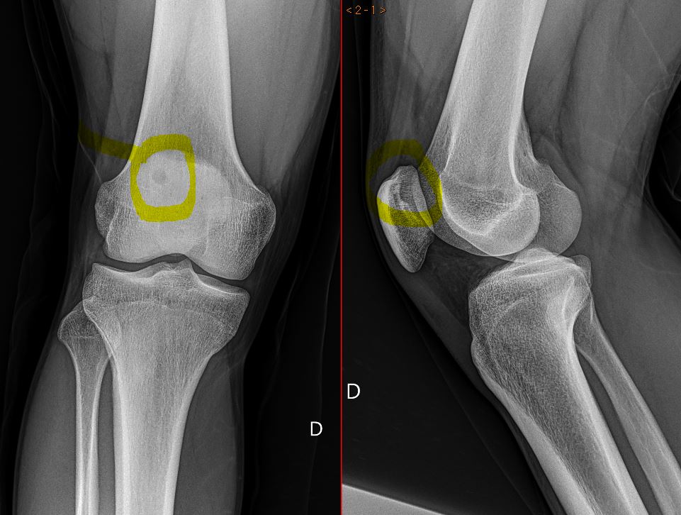
24/1/2024. Painfull #knee #MRI: 1⃣Symptomatic dorsal patella defect or 2⃣postraumatic ostechondral lesion? According to the morphology and location of the lesion, one of the diagnosis can be suggested. 🤔 Share your thoughts! More MRI sequences and insights coming soon. 🚀

0
11
86
@handoshera I just describe the finding, specially if there is no other pathology in the MRI. I doubt if, in this example, it explains the knee pain. Thanks you!
1
0
4
DIAGNOSIS ⛔️ - Os acromiale 🦴with degenerative changes in the synchondrosis.


2
11
79

RT @themskarchive: @pepelermarx @drwillyjara @JM_Zulueta_O @SermeMsk @ESSRmsk DIAGNOSIS - 2⃣ Type III tripartite patella with degenerative…
0
7
0
RT @themskarchive: 5/4/2024. 🟠 23yo male presents to ED with left #knee 🦵contusion. #XRAY reveals: 1⃣ Superolateral patellar fracture, tw…
0
15
0
9/1/2025. Bipartite patella Type I 🦴: a congenital variant with an unfused inferior patellar pole. On 🩻, look for a distinct fragment with smooth margins; MRI reveals a fibrocartilaginous bridge with mild degenerative changes. #Radiology #Ortho #BoneHealth


0
8
43

1
0
5