
Histology
@histocasino
Followers
3K
Following
484
Statuses
3K
Bet wisely in the Histo Casino if you want to win big. I am Gerardo Ochoa-Vargas, MD. My main Twitter is @gerardo8av
Joined February 2010
Children enjoy microscopes, as they can gaze into a hidden world! #BestToyEver #WinterIsComing #SantaIsComing

5
37
86
Not Histology, but still worth watching, to see the power of a scanning electronic microscope #SEM made available by @IBMResearch @IBM
0
0
0
RT @sciam: Google scientists have modelled a fragment of the human brain at nanoscale resolution, all 150 million connections https://t.co/…
0
82
0
RT @DevinMynett: @HopeRising19 Angus Dalgleish was allegedly trying to sell a vaccine to compete with Pfizer/Astra Zeneca that he had share…
0
1
0
Histology students should identify the Reinke crystals (or the spaces left by them) in the Leydig cells shown in the third panel
0
0
5


Desmosomes (maculae adherentes) have an attachment plaque made of desmoplakin and plakoglobin, to which intermediate filaments (tonofilaments) are anchored
0
0
2
The author meant osteoclasts, which are also multinucleated. Osteoblasts are mononuclear. Also remember that osteoclasts are not macrophages.

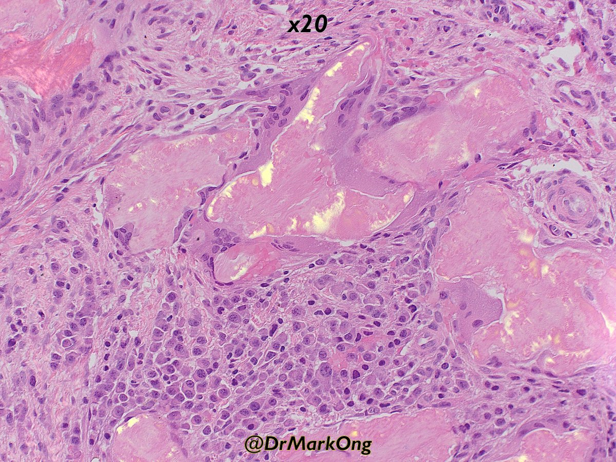
Thank you all. This was indeed a plasma cell neoplasm with abundant amyloid. What looks like cortical bone is amyloid. The multinucleate cells are foreign body type giant cells, not osteoblasts. This is better appreciated in this picture 👇🏻

0
0
2
Indeed: abundant amyloid may elicit birrefringence even if Congo Red is not used. It is always an interesting experience to try to detect it without the use of that dye, in the HE stained tissue. Fantastic image!
If there is enough amyloid, you may not need Congo Red to demonstrate birefringence, as demonstrated below on the H&E. The chemical(s) in Congo Red intercalate between amyloid fibrils, accentuating the inherent birefringence of the amyloid.

0
1
5
Histology students always expect mast cells to be stained with stains that yield metachromasia, as toluidine blue. With HE, notice the contrast with the neighbouring cells (cytoplasm is more red, brighter) and the finely granular pattern of the cytoplasm. Practice!

I'm often found in secretory meningiomas. What kind of cell am I? (If you get this right, you've MASTered an interesting little aspect of pathology.) #pathology #neuropath #PathTwitter

0
0
9
The blue dense areas are most likely corpora albicantia (plural of corpus albicans), scars formed where corpora lutea (plural of corpus luteum) used to exist

2/🧵 Cross section of a human female ovary. That is what makes a human female, a woman 😀

1
0
8
RT @TheKoshurDoc: 2/🧵 Cross section of a human female ovary. That is what makes a human female, a woman 😀
0
3
0
RT @TheKoshurDoc: Cross Section of a human female ovary. Can you name the major structures seen? Isn’t it beautiful? #histology #MedEd #M…
0
5
0
RT @FdeCastroS: I've just known the recent death of Don Miguel #MarínPadilla,great neuroanatomist,MD by @CanalUGR,formed at @InstitutoCajal…
0
9
0
RT @TheEconomist: When the Human Cell Atlas began, no one knew how many types of cell there were. The answer turns out to be: “a lot more t…
0
9
0
Gorgeous expression of desmosomes joining keratinocytes

What are these cells & how do you know? Answer: #Pathology #dermpath #dermatology #dermatologia #dermtwitter #pathologists #pathTwitter #histology

0
3
13