
SAR DFP Benign Biliary Pathology
@SARBiliaryDFP
Followers
318
Following
50
Statuses
125
Official Account of the Society of Abdominal Radiology (SAR) Benign Biliary Pathology Disease Focused Panel
Joined March 2021
@RachitaKhot @UVARadiology Small, central signal void in the CBD on the axial plane without correlation on different T2WI planes. This finding ion fast spin-echo or gradient-echo sequences is due to their sensitivity to motion and flow. Diagnosis: Pseudo-filling defect due to flow artifact
0
4
7
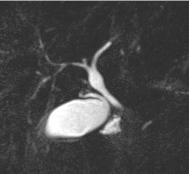
Multifocal cystic dilatation of the intrahepatic bile ducts with visible enhancing central portal radicle. •Diagnosis: Caroli’s Disease (Type 5 choledochal cyst)
0
1
2
Challenge yourself with another biliary case: Case courtesy of Daniel R. Phadke from UVA Radiology @UVARadiology 44 yo woman with history of epigastric and left upper quadrant pain
1
1
9
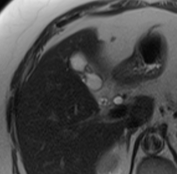
Diagnosis: Segmental Adenomyomatosis Imaging findings: Hourglass configuration of the gallbladder with cystic changes in the wall. It occurs from hyperplasia of mucosal epithelium which invaginates into the muscularis, forming Rokitansky-Aschoffsinuses.
Challenge yourself with another biliary case: Case courtesy of Daniel R. Phadke from UVA Radiology @UVARadiology 70 yo woman who presents for follow-up of incidentally detected cystic renal mass. No RUQ symptoms.
1
1
9

Challenge yourself with another biliary case: Case courtesy of Daniel R. Phadke from UVA Radiology @UVARadiology 70 yo woman who presents for follow-up of incidentally detected cystic renal mass. No RUQ symptoms.
3
6
14
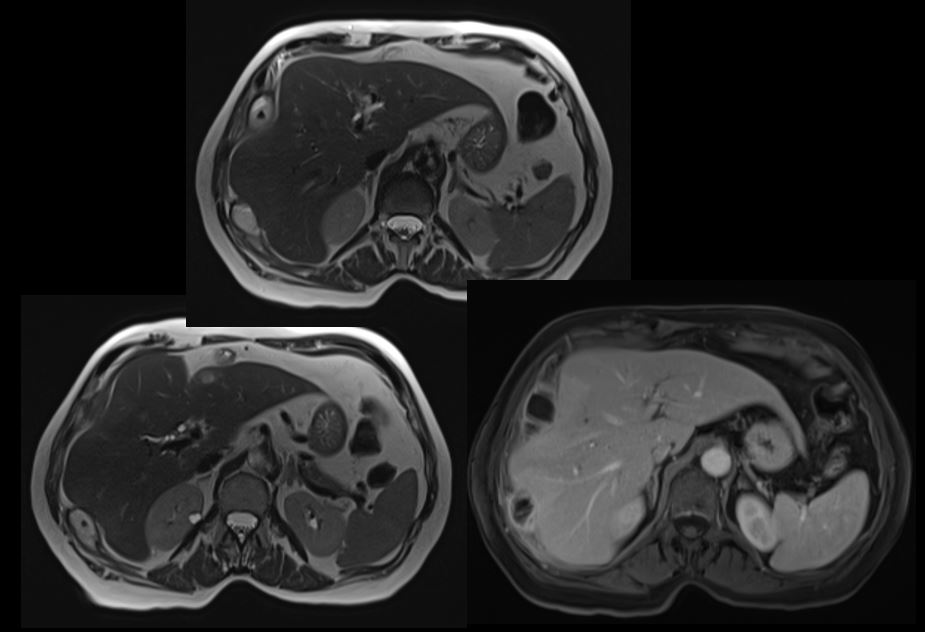
Diagnosis: Multiple dropped gallstones. Imaging findings: Multiple areas of inflammation and collections around T2-hypointense foci (corresponding to dropped gallstones).
Challenge yourself with another biliary case: Case courtesy of Malak Itani MD @ItaniMalak from Mallinckrodt @MIRimaging 78 y.o. woman with abdominal pain 6 months after cholecystectomy
0
3
8
@ItaniMalak Answer: Imaging findings: Numerous areas of inflammation and developing collections around T2-hypointense foci (corresponding to dropped gallstones). •Diagnosis: Multifocal dropped gallstones
Challenge yourself with another biliary case: Case courtesy of Malak Itani MD @ItaniMalak from Mallinckrodt @MIRimaging 78 y.o. woman with abdominal pain 6 months after cholecystectomy
0
0
3
Challenge yourself with another biliary case: Case courtesy of Malak Itani MD @ItaniMalak from Mallinckrodt @MIRimaging 78 y.o. woman with abdominal pain 6 months after cholecystectomy
4
4
15
Diagnosis: Cholecystoduodenal fistula. Imaging findings: Chronic cholecystoduodenal fistula with an impacted gallstone.
Challenge yourself with another biliary case: Case courtesy of Malak Itani MD @ItaniMalak from Mallinckrodt @MIRimaging 63 y.o. woman with cirrhosis

0
3
5
Diagnosis: Cholecystoduodenal fistula @ItaniMalak Imaging findings: Chronic cholecystoduodenal fistula with an impacted gallstone. •
0
0
1
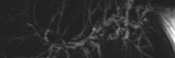
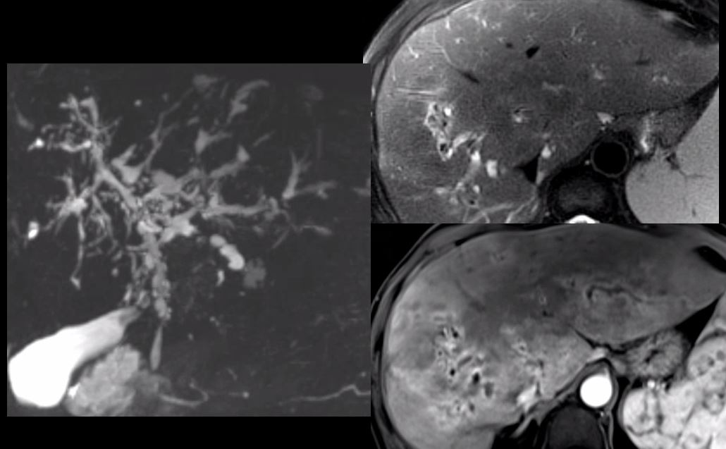
Primary Sclerosing Cholangitis with active infection case courtesy of @andersonmark27 and MGH radiology @mghradchiefs Imaging findings: multifocal intrahepatic and extrahepatic stricturing with bile duct thickening, enhancement, and peribiliary inflammation
Test Yourself! A 55-yo man with history of ulcerative colitis presents with fever, right upper quadrant pain, and LFT abnormalities. MRCP MIP image, axial T2-weighted image with fat sat, and axial T1-weighted post-contrast image with fat sat. Answer will be posted in a week.
0
1
8
RT @SARBiliaryDFP: Test Yourself! A 55-yo man with history of ulcerative colitis presents with fever, right upper quadrant pain, and LFT ab…
0
8
0
Test Yourself! A 55-yo man with history of ulcerative colitis presents with fever, right upper quadrant pain, and LFT abnormalities. MRCP MIP image, axial T2-weighted image with fat sat, and axial T1-weighted post-contrast image with fat sat. Answer will be posted in a week.
1
8
15
Another excellent case from our Benign Biliary DFP. Many more to come! Follow us @SARBiliaryDFP @SocietyAbdRad


0
3
5
Check out the first of many coming education case series from the Benign Biliary DFP @SocietyAbdRad


0
4
7
RT @Abdominal_Rad: Graphic abstract published 5/18/2022 #abdradJ #radiology #imaging #abdominalimaging Secondary sclerosing cholangitis:…
0
13
0