

Brookes Bioimaging
@OBBU_microscopy
Followers
128
Following
32
Statuses
60
RT @DrLouiseCHughes: Because #fridayfeeling i am also going to post the abstract art stemming from today's #microscopyadvent of lily pollen…
0
11
0
RT @DrLouiseCHughes: 2/12 Close up of a lily pollen grain captured with SEM. #microscopyadvent #microscopyart #sciart @MicroscopySoc @obu_b…
0
15
0
RT @DrLouiseCHughes: It is that time of year again! #microscopyadvent #1stdecember ##AdventCalendar SEM of Iris pollen grains #sciart @obu_…
0
9
0
RT @DrLouiseCHughes: SEM image (after some editing) of willow pollen, combining BSE and SE signals. #sciart #biology @obu_bms @oxford_brook…
0
8
0

3D reconstruction of a Golgi apparatus from Arabidopsis root cells. #biologyweek #sciart @brookeshls @zeiss_micro @OBBU_microscopy #3DEM

0
0
1
Inside a root looking at the nuclei. 3D SEM data for VR #virtualreality #realdata #biologyweek #sciart @zeiss_micro @brookeshls

0
0
0
I have won the @RoyalSocBio Science Communication Award for an established researcher! #thrilled #scicomm
0
2
0
RT @royalsociety: Our very special collection of the life & works of Robert Hooke - Under the microscope https://t.…
0
48
0
Incredible book and images.

#RobertHooke died on this day in 1703. We're celebrating his greatest work #Micrographia for #WorldBookDay

0
1
1
Some of the work done here at OBBU. #brookeshls
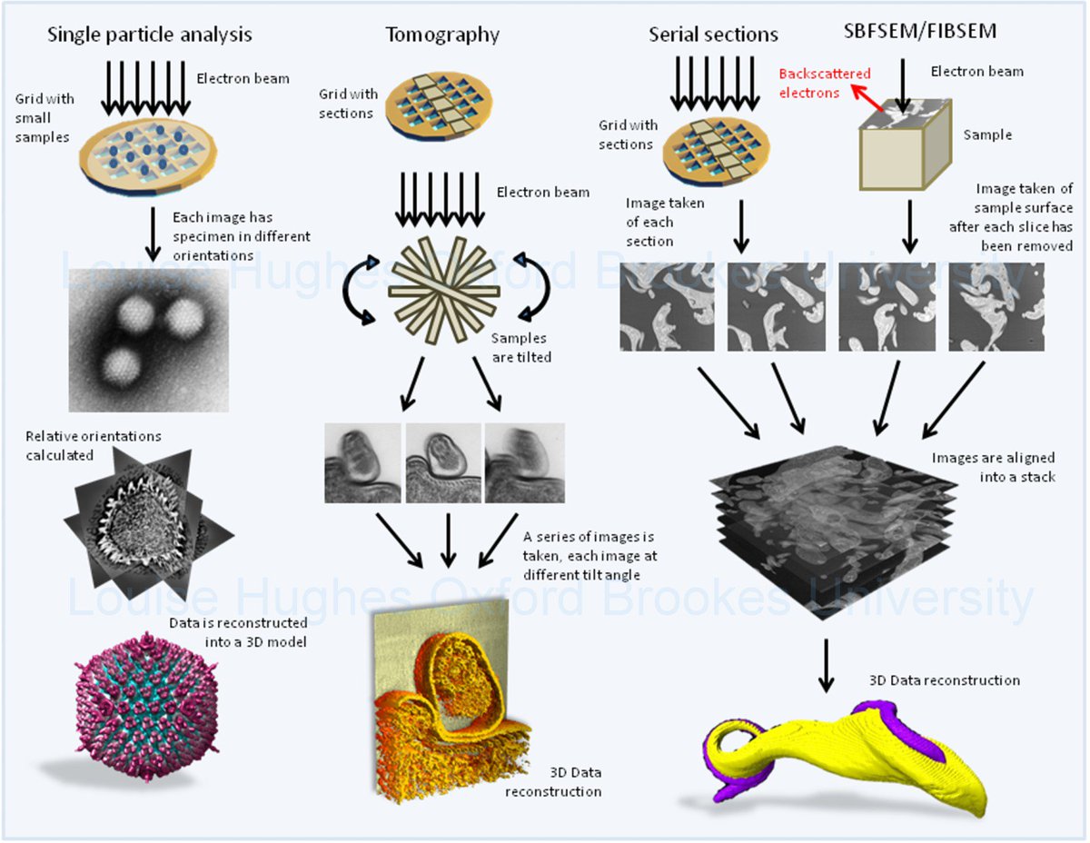
How to get 3D data using electron microscopy. Diagram showing some of the different techniques. #sciart
0
3
1
Microscopy club for BMS undergraduates every other Wednesday afternoon!
Flea mite imaged with SEM. Taken during @OBBU_microscopy microscopy club for undergrads. #brookeshls #sciart

0
0
0
Undergrad teaching this pm!
TEM prac. with undergrads this pm. Image of plant Golgi bodies. #brookeshls #sciart @HawesChris @jakerich101

0
0
0
This was captured using serial block face scanning electron microscopy! Beautiful data. @zeiss_micro
#microscopyadvent day 18. 3D data - stoma and the surrounding guard cells in a leaf @zeiss_micro #sciart #brookeshls

0
0
1
RT @zeiss_micro: #ZEISS #Airyscan: confocal #microscopy with improved signal-to-noise ratio & super-resolution @naturemethods
https://t.co/…
0
2
0
Transmission electron microscopy image. Good to see some traditional EM going on. :)
#microscopyadvent day 17. Another Golgi body. This is a coloured TEM image of an animal cell. #sciart #brookeshls

0
1
0
RT @hls_research: Ever wondered how a Scanning Electron Microscope works? Prof @HawesChris explains @OBBU_microscop…
0
6
0

0
0
0
RT @DrLouiseCHughes: #microscopyadvent day 14. SEM image of the tip of a Drosophila proboscis, fruit fly mouthparts. #sciart #brookeshls ht…
0
7
0
RT @DrLouiseCHughes: How a SEM works as described by @HawesChris showing microscopes in our bioimaging unit as well as some of my data. htt…
0
6
0