

MRI-ESSENTIALS
@EssentialsMri
Followers
6K
Following
157
Statuses
217
Wolfgang Fischer, MSK radiologist. I publish the App "MRI-ESSENTIALS" (iOS, Android) and I am happy to get your feedback.
Augsburg
Joined December 2019
Skiing on New Year's Day: Fracture of the lateral tibial plateau with interposition of the lateral meniscus (arrowhead). #mskrad #orthotwitter #radres #radiology
2
1
16
A small crown for the holy three kings. Thanks to the hydroxyapatite deposit! #mskrad #orthotwitter #radres #radiology

1
0
11
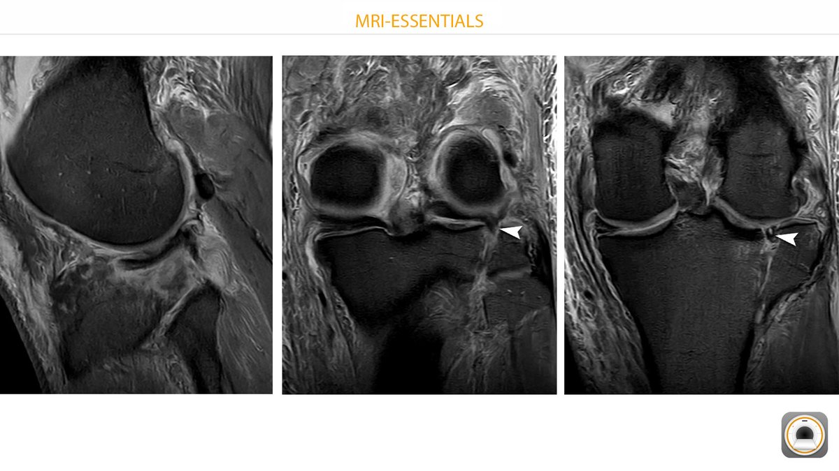
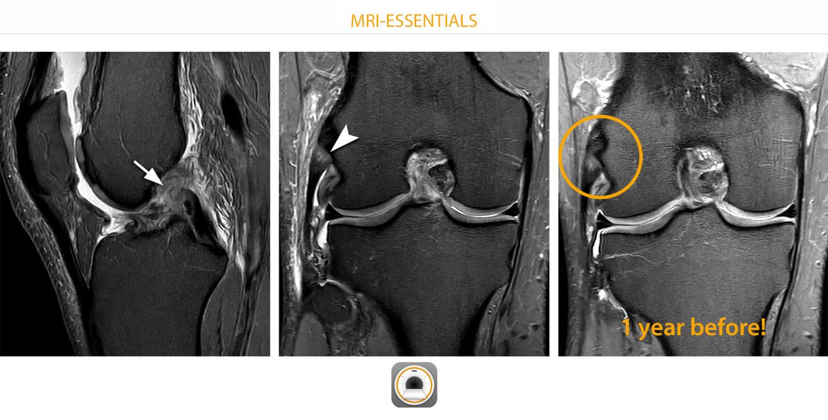
LCL knee - always a pitfall! Left: Ski injury on New Year's Day, tear of the ACL (arrow). Center: A partial tear of the lateral collateral ligament could also be suspected (arrowhead). But: It looked exactly the same a year ago! #mskrad #orthotwitter #radres #radiology
1
3
26
@dottorsanna Hello Nicola. Thanks for the message. Indeed I forgot to label it in the hip section although it is visible in the axial and coronal plane. Will fix this issue with the next update (probable february). Thanks and Happy New Year 2024!
0
0
0
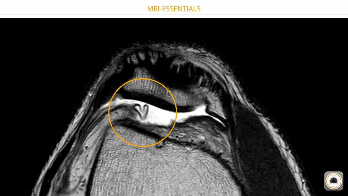
The suprapatellar plica and MRI-ESSENTIALS wish you a Happy New Year 2024! 🙂 #mskrad #orthotwitter #radres #radiology
0
1
14
#RSNA23 Just stop by and visit us at booth 1303, South hall. I´ll show you the new web version of MRI-ESSENTIALS: #mskrad #orthotwitter #radres #radiology

1
2
15
If you are attending RSNA in Chicago, come visit me at booth 1303 in the educators row (South Hall). I´ll show you the new WEB VERSION of MRI-ESSENTIALS! #mskrad #orthotwitter #radres #radiology

0
1
5
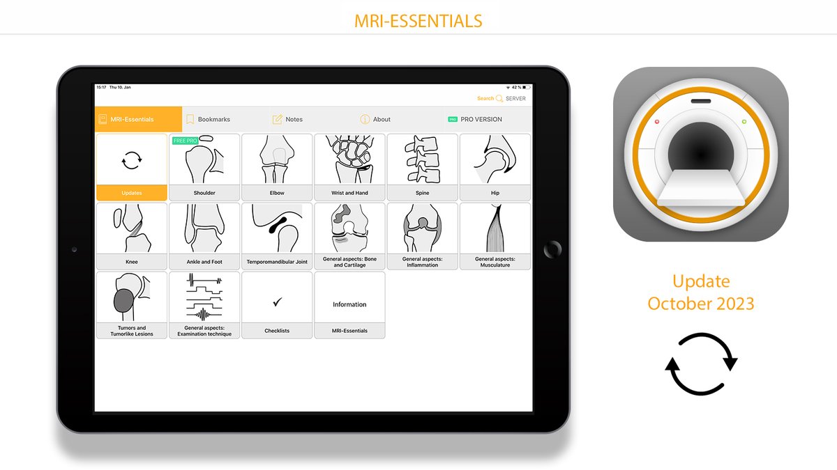
Quick info for users of the app MRI-ESSENTIALS: I have just released a new update. Content from current literature has been incorporated in the chapters Shoulder, wrist and hand, Spine and SIJ. The next update is planned for November. #mskrad #orthotwitter #radres #radiology
0
0
9
@GSERRANOB_MSK Nice video. However, it seems that biceps instability may also occur without tear of the pulley - just insufficiency of intact structures. This makes it difficult for us to exclude biceps instability. Eg. #mskrad #orthotwitter #radres #radiology
1
0
1
Pitfall: The arrow does not show the root of the medial meniscus but a strong meniscotibial ligament (fibrotic due to torn root???)! The root would be located anterior to the PCL, as indicated with the arrowhead. #mskrad #orthotwitter #radres #radiology

0
2
12
Rare case of a discoid MEDIAL meniscus, 14 yo boy. Note the significant degeneration with formation of a ganglion cyst causing pain. #mskrad #orthotwitter #radres #radiology

0
5
37
Instability of the lateral meniscus. Left: Acute knee locking, examination in flexion! The next day: normal meniscal position. 3 years ago, the attachments of the post. horn were already missing (Wrisberg type of the lateral meniscus)! #mskrad #orthotwitter #radres #radiology

0
15
57
Tear of the superficial flexor tendon (arrow) due to chronic inflammation in a 57 year-old man with rheumatoid arthritis. Synovitis in the MCP joints and impressive synovitis along the tendon sheath. (Bottom right: with contrast) #mskrad #orthotwitter #radres #radiology

0
9
39
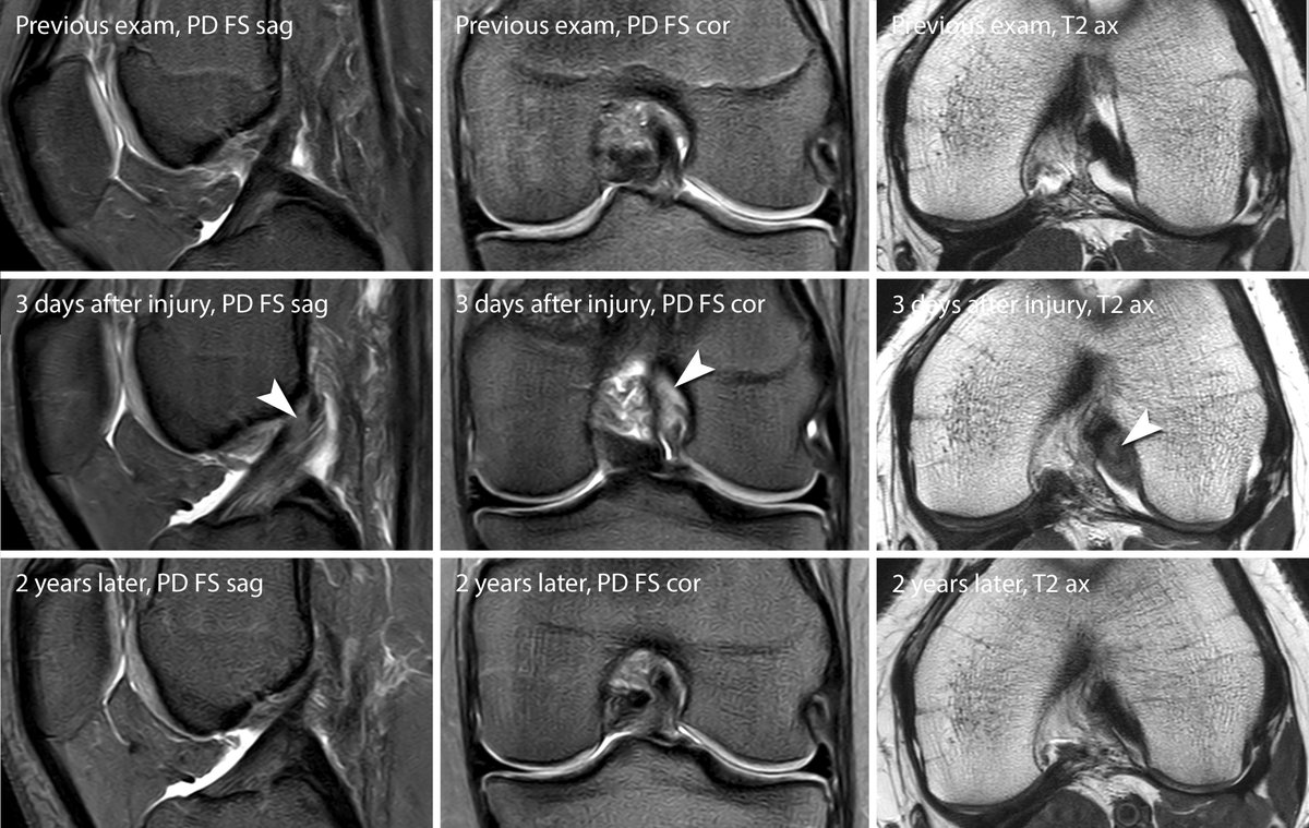
Partial tears ACL tears: always difficult. Often we do not know if the diagnosis was correct (conservative therapy). In this case, comparison with an older and later exam demonstrates the injury at the origin of the posterolateral bundle. #mskrad #orthotwitter #radres #radiology
2
8
52
Together with Kay G. Hermann we developed a module for the BERLIN CASE VIEWER on the topic of meniscus tears. Great interactive teaching tool! #mskrad #orthotwitter #radres #radiology #berlincaseviewer
0
1
10
I have published a new update for MRI-ESSENTIALS. Focus 1: rotator interval, bicpes pulley. Focus 2: elbow instability. Feedback is always welcome! #mskrad #orthotwitter #radres #radiology

1
4
30
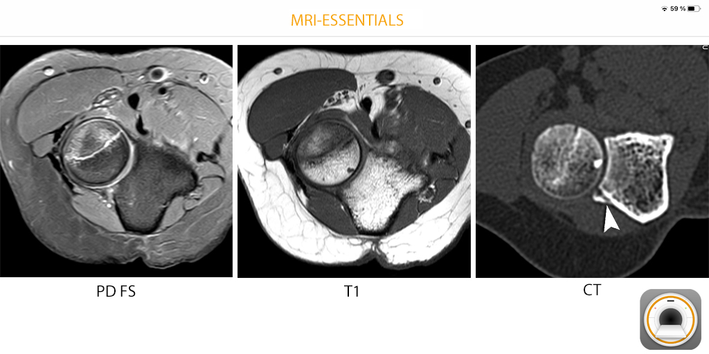
Obvious fracture of the radial head. The bony avulsion of the annular ligament was missed on MRI. The small fragment is clearly visible on CT (arrowhead). #mskrad #orthotwitter #radres #radiology
2
3
21
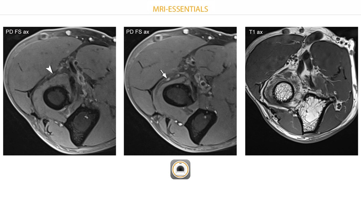
Radial tunnel syndrome. Note the atrophy of the supinator muscle in T1. The deep branch of the radial nerve is unremarkable before entering the supinator (arrowhead) and shows neuritis slightly more distally (arrow). #mskrad #orthotwitter #radres #radiology
0
10
46
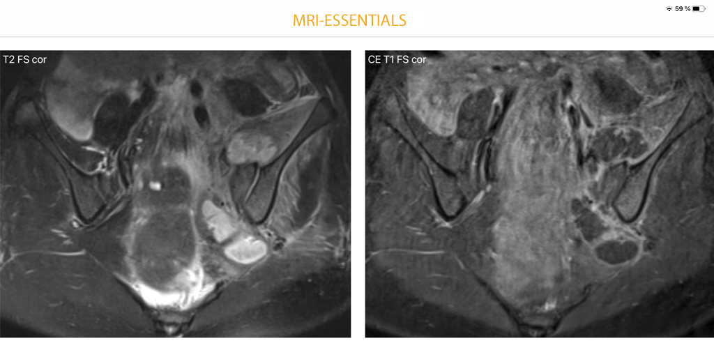
Bacterial sacroiliitis with diffuse contrast enhancement around the left sacroiliac joint and extensive abscesses. Unremarkable right SIJ. Even without abscess formation, infection would have to be suspected because of the asymmetry. #mskrad #orthotwitter #radres #radiology
0
6
17