

Dr Razi
@DrRazi4
Followers
4K
Following
10K
Statuses
8K
ECG collector
Kuala Lumpur Malaysia
Joined February 2018
@suricardio @amreshgul @baramink @CardioBeat_ @chi_no_usagi @DrRajeshG1 @ecgandrhythmRoe @FibrilloFlutter @makkyecg Great case; thanks for sharing👏👏
0
0
1


@suricardio @amreshgul @baramink @CardioBeat_ @chi_no_usagi @ecgandrhythmRoe @FibrilloFlutter @Ahmedata7777 OMI signs detected ‼️‼️🚑🚑 #ecg shows: 1. STE with hyperacute TW in Inferior Leads 2. STE in III>II 3. Reciprocal STD aVL & Lead 1 4. Max STD V1-V4 ⏭️Posterior OMI - Confirmed by Mirror #ecg 5. No lateral involvement 🔺Acute Inferior Posterior OMI (RCA occlusion)


2
4
9


Good catch👏

@ecgandrhythmRoe @EcgOxford @DrRazi4 @PendellM @RobertHermanMD @Arron_Pearce_ @ECGwithReid @EM_RESUS Answer: anterior OMI with cardiogenic shock 🚨 Coronary angio showed severe 3-vessel disease with occlusion of pLAD, subtotal occlusion of mid CX, and CTO of pRCA and DA1. Despite quick and easy revascularization of pLAD and CX lesion, Impella insertion was needed.


0
0
1
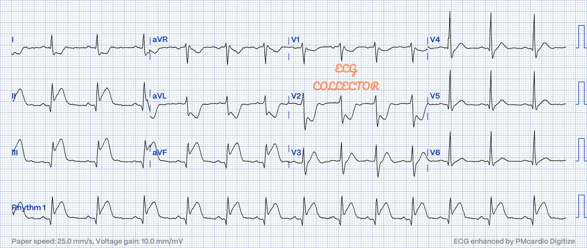
Young gentleman at 39 y.o with chest pain; How many areas are involved here❓🤔 @PMcardioApp
@LITFLblog
#Cardiology
#CardioTwitter
#CardioEd
#Medicalstudent
#Medtwitter
20
13
56
@KoshalMeena2001 @ecgandrhythmRoe @Frances98392343 @LeonrMail @Ecgloverr @koggelnoggel @EM_RESUS @EcgsOnly @EcgOxford @EkgHacks @ECGWeekly What type of inflammation ?
2
0
0
Great👏

Are you using the @PMcardioApp for subtle #OMI detection? Proportionality matters—here is how verifying the digitized waveform in poor-quality #ECG images can prevent false positives. Read our @JElectrocardiol Editorial on @Marco__Biasin's pneumothorax case.

0
1
10
#ecg shows NCT HR ~170bpm: 1. Long RP (RP>PR) 2. 1:1 AV relationship 3. (+) P wave inferior leads - excluded AVNRT/AVRT 4. (+) P wave in aVR - excluded ST 5. (-) P wave in V1-V6 🔺Focal Atrial Tachycardia (RA focus ⏭️🤔possible Tricuspid Annulus)


46 years old man with sudden onset palpitations. Vital signs normal Labs normal What’s the rhythm? Notice the 1st image Increased to 20 mm/mV.


1
9
36
Always do the rhythm strip while giving Adenosine to see the clear response in view of its short half life

Responses of Narrow Complex Tachycardias to adenosine ESC guidelines 3 different effects on Atrial Tachycardia

3
7
21
@KoshalMeena2001 @ecgandrhythmRoe @Frances98392343 @LeonrMail @Ecgloverr @koggelnoggel @EM_RESUS @EcgsOnly @EcgOxford @EkgHacks @ECGWeekly ST usually responds to various non-cardiac causes such as anaemia, thyroid, anxiety, electrolytes and etc. The pain also can induce ST; If you cannot find any of the above causes; can start with managing the pain first with adequate analgesia.
1
0
2
@MubarakAlhatemi @EM_RESUS @The_Nanashi_O @ecgrhythms @syamkumarmd @adribaran #ecg shows NCT HR ~170bpm: 1. Long RP (RP>PR) 2. 1:1 AV relationship 3. (+) P wave inferior leads - excluded AVNRT/AVRT 4. (+) P wave in aVR - excluded ST 5. (-) P wave in V1-V6 🔺Focal Atrial Tachycardia (RA focus ⏭️🤔possible Tricuspid Annulus)


0
1
12
@KoshalMeena2001 @LeonrMail @ecgandrhythmRoe @Ecgloverr @koggelnoggel @EM_RESUS @EcgsOnly @Frances98392343 @EcgOxford @EkgHacks @ECGWeekly Sinus tachycardia..no OMI signs detected. Usually, ACS will not come with tachycardia unless they are going to/in cardiogenic shock.
3
0
4
@LeonrMail @ecgandrhythmRoe @Ecgloverr @koggelnoggel @EM_RESUS @EcgsOnly @Frances98392343 @EcgOxford @EkgHacks @ECGWeekly This is just a minor elevation: it fits Wallen's
1
0
6