

Ximena Wortsman
@xworts
Followers
534
Following
3K
Statuses
1K
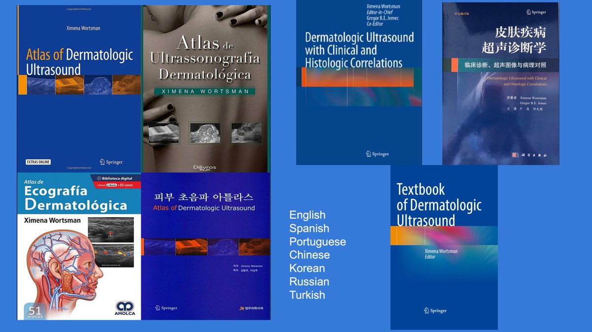
Radiologist, Ranked number 1 worldwide in publications and citations on Dermatologic Ultrasound. 3 books/262 publications. Derm US @sonoskin
Joined February 2009
RT @TheLancet: Hidradenitis Suppurativa is a chronic inflammatory disease that has a profound impact on patients' quality of life. Discove…
0
44
0
🔥🔥🔥 Just published!!! First report on ultrasound pattern of congenital smooth muscle hamartoma (CSMH) of the skin, representing the most frequent type of smooth muscle hamartoma. Ultrasonographic features at 24 MHz and 70 MHz can support clinical diagnosis and follow-up of CSMH, which may potentially avoid biopsies. More info at: Meza V, Aranibar L, Wortsman X. Ultrasound Pattern of Congenital Smooth Muscle Hamartoma of the Skin. J Ultrasound Med. 2024 Dec 18. doi: 10.1002/jum.16636. Epub ahead of print. PMID: 39692078. Link: @xworts @sonoskin @AIUMultrasound

0
2
9
I’m honored to have been invited as an international speaker at CILAD 2024, the Ibero-American Congress of Dermatology, which took place in the beautiful city of Cartagena, Colombia. Over 4,000 attendees participated in this congress. I presented on several topics related to dermatologic ultrasound. It was a fantastic opportunity to learn, teach, and reconnect with old friends while enjoying the warmth of Colombia. #dermatologicultrasound #cartagena #hidradenitissuppurativa #ultrasound #nails #dermatology #fillers @xworts @sonoskin @CILADderma
0
1
5
Hidradenitis suppurativa has greatly benefited from the insights of color Doppler ultrasound. Ultrasonography has helped prove the follicular link of this disease and has ruled out the primary involvement of the apocrine glands, which, in the old days, was supposedly the cause of the disease. Importantly, ultrasound can detect subclinical anatomical information in HS that cannot be deducted from the clinical examination. Moreover, high-frequency (≥15 MHz) and ultra-high-frequency (≥50 MHz) ultrasound present a much higher axial spatial resolution compared to magnetic resonance imaging. Ultrasound allows us to detect better subclinical cutaneous anatomical abnormalities and, therefore, arrive earlier and more accurately at diagnosis and staging. Ultrasonographic diagnostic criteria can discriminate HS from other clinical simulators, which is also critical in diagnosing mild and moderate stages and is relevant for the severe stages. This imaging technique supports the severity (mSOS-HS) and activity (US-HSA) scorings more accurately, which can help assess the actual stage of the disease. This is important to decide the type of treatment and to perform a more objective follow-up of the patients. Magnetic resonance imaging has been reported as helpful in diagnosing deep perianal tunnels; however, it presents a lower axial spatial resolution compared with high and ultra-high frequency ultrasound. Nowadays, there is solid evidence of the usefulness of ultrasound in HS, which implies that it is a game-changer and should be recommended as the first-choice imaging technique and a standard of care tool for HS patients. More info at; Wortsman X. Update on the role of color Doppler ultrasound in hidradenitis suppurativa: a game-changer. Ital J Dermatol Venerol. 2024 Nov 19. doi: 10.23736/S2784-8671.24.08025-3. Epub ahead of print. PMID: 39560343. @xworts @sonoskin

0
6
15
I am grateful to have received two formal recognitions from the Brazilian Society of Ultrasound (SBUS) and the Post Dermus in Sao Paulo, Brazil. The SBUS 2024 annual meeting and the Post Dermus had full days of interesting dermatologic ultrasound lectures. In the Post Dermus, there was also a hands-on session. It was an excellent opportunity to meet with great old and new colleagues and friends working in the field and enjoy the Brazilian warmth. Special thanks to Dr. Giselle Goes and Dr Paula Colpas #dermatologicultrasound @xworts @sonoskin


1
1
9
🔥🔥🔥 Don’t miss it!!! This review, which is Open Access and just published, practically analyzes the main ultrasonographic features of the most common types of skin cancers and the performance of locoregional staging according to the literature. It is illustrated with state-of-the-art clinical and ultrasonographic correlations. The most common types of skin cancer show recognizable ultrasonographic patterns. Among the current radiological imaging techniques, ultrasound has the highest axial spatial resolution. Compared to other imaging techniques used in dermatology, ultrasound has the great advantage of penetrating the soft tissues thoroughly, which allows us to detect and identify the most common types of skin cancer, including both the primary tumor and its locoregional metastases. Link to Article: @xworts @sonoskin
1
13
44
🔥🔥🔥This case-control study evaluates the effectiveness of ultrasound (greyscale, color Doppler, and shear wave elastography (SWE) in distinguishing between active and inactive morphea lesions, aiming to improve the accuracy of morphea management. Our results indicated that the final radiologist's assessments based on Wortsman's criteria and increased hypodermal echogenicity achieved high sensitivities (0.94) and specificities (0.88). Our findings support using grayscale US, color Doppler, and SWE for a precise and thorough assessment of morphea activity. More info at: Etesami I, Azizi N, Sabrinejad R, Montazeri S, Kamyab K, Nasimi M, Mahmoudi H, Khorasanizadeh F, Wortsman X. Sonographic Skin Features and Shear Wave Elastography in Distinguishing Active from Inactive Morphea Lesions: A Case-control Study. J Am Acad Dermatol. 2024 Sep 21:S0190-9622(24)02877-9. doi: 10.1016/j.jaad.2024.09.026. PMID: 39313033. @xworts @sonoskin
0
2
5
🔥🔥🔥This article just published shows the ultrasonographic pattern of the new generation of polymethylmethacrylate (PMMA), which is currently used in aesthetic procedures in some countries. There are some differences in high-frequency ultrasound compared with the old PMMA, but at ultra-high frequencies, the patterns are similar between the new and old ones. Recognizing the pattern on ultrasound is essential since this is the first-choice imaging technique for detecting and discriminating cosmetic fillers. More information at: Sigrist R, Fassina M, Wortsman X. Ultrasonographic Pattern of a New Generation of Polymethylmethacrylate at High-Frequency and Ultra-High Frequency. Dermatol Surg. 2024 Oct 3. doi: 10.1097/DSS.0000000000004422. Epub ahead of print. PMID: 39361534. @xworts @sonoskin

0
3
5
I am grateful and glad to have been a speaker at #EADV2024 in #Amsterdam. Our #dermatologicultrasound course was sold out, and the nail session had a full house. Besides, I presented our work on facial and submandibular hidradenitis suppurativa in the EHSF session, which was very well-received. Special thanks to Dr. Fernando Alfageme, Dr. Peter Velthuis, and Ms. Egle Urbonaite for greatly supporting the organization of the dermatologic ultrasound course. @xworts @sonoskin #dermatologicultrasound #skinultrasound #nailultrasound
@eadv
0
1
7
Glad to be lecturing about dermatologic ultrasound at the University Hospital Erasmus in Rotterdam, The Netherlands. Special thanks to Dr. Peter Velthuis, Dr. Leonie Schelke, and Dr. Sara Koshaw.


0
1
6
Don´t miss this article, which was just published in JEADV ! This review emphasizes the importance of imaging in nail diseases. It describes the benefits and limitations of the most commonly used imaging techniques and their level of evidence. The role of ultrasound in supporting the diagnosis of nail diseases is relevant and covers more pathologies compared to MRI, OCT, LC-OCT, and Confocal Microscopy. Moreover, it allows us to observe all the parts of the nail unit without penetration issues. More information: Sechi A, Wortsman X, Tosti A, Iorizzo M. Advances in image-based diagnosis of nail disorders. J Eur Acad Dermatol Venereol. 2024 Sep 4. doi: 10.1111/jdv.20309. Epub ahead of print. PMID: 39230323. Link to article: @xworts @sonoskin

0
5
9
I was pleased to lecture at the Nail Society of India on August 28, 2024, about the “Role of Ultrasound in Nail Pathologies.” I would like to thank Dr. Chander Grover for the kind invitation. This activity was a CME for Indian dermatologists. #nailultrasound #nails #dermatologicultrasound @xworts @sonoskin

0
2
7