

Sameer Raniga
@samrad77
Followers
16,709
Following
4,395
Media
3,075
Statuses
9,620
Radiologist. Trauma and Emergency Radiology. One view is NO view. #radtwitter | #FOAMrad | #radres | #radEd | #radiology | #EmergencyRad
Muscat, Oman
Joined March 2013
Don't wanna be here?
Send us removal request.
Explore trending content on Musk Viewer
Cheney
• 362216 Tweets
Eagles
• 228072 Tweets
Packers
• 131884 Tweets
ENGFA ACTRESS 100M
• 102564 Tweets
श्री गणेश
• 101074 Tweets
Ganesh Chaturthi
• 87524 Tweets
Ganesh Chaturthi
• 87524 Tweets
#ATIPASHOPxCHARLOTTESNACK
• 82202 Tweets
Saquon
• 74596 Tweets
Perú
• 73840 Tweets
गणपति बप्पा
• 73341 Tweets
Jalen
• 57356 Tweets
Green Bay
• 37672 Tweets
भगवान गणेश
• 35959 Tweets
#キントレ
• 32975 Tweets
Jordan Love
• 30718 Tweets
#二度と撮れない画像を貼れ
• 27534 Tweets
Duke
• 25916 Tweets
#GanpatiBappaMorya
• 25546 Tweets
श्री सचिन पायलट
• 20349 Tweets
Lucho
• 14854 Tweets
#音泉祭り
• 14253 Tweets
ビッグラン
• 14188 Tweets
Jayden Reed
• 13860 Tweets
Party Love AndaLookkaew
• 13740 Tweets
JustinOn NCAA100Kickoff
• 13304 Tweets
イコラブ
• 12510 Tweets
#2024TRUSTY_IN_BANGKOK
• 10360 Tweets


Pinned Tweet

This will be my pinned tweet. Till the end of time.
Let's demyth the contrast induced nephropathy (better contrast associated acute kidney injury).
Thank you
@DGlaucomflecken

4
15
110


A group of Radiologists avoiding direct patient interaction 😀
33
182
1K
The first CT scanner was created by British engineer Godfrey Hounsfield in 1972.
ER physicians before 1972:
55
241
954
ER physician sending patients for imaging during my on-call: 😅
23
111
836
How NOT to miss a compression fracture of the vertebral body on CT?
Look for the following features: 5Ds
1. Dense (sclerotic) # line
2. Depression- wedging
3. Deformity- end plates (depression)
4. Deformity- buckling or cortical step
5. Discontinuity- cortex (corners)


7
152
609
A picture is worth 1,000 words!
Reference: New Bone Formation in Axial Spondyloarthritis: A Review. Rofo. 2023 Nov 9. English. doi: 10.1055/a-2193-1970. Epub ahead of print. PMID: 37944938.

4
147
589
Urologists rightly advise that frequently emptying the bladder can save your life from the risk of stones... This is an example...
😅
11
145
588
Was driving home today when I thought I saw a Spy Balloon, until I realized it was Birdshit on my Windshield 😅
Lesson: One view is NO view.. 🫢

25
37
544
That one Radiologist in your department who never retires… 😀
9
54
501
Radiologist and pathologist in tumor board when the others are discussing management plan..
10
84
495
A beautiful diagram showing spectrum CT findings in Interstitial fibrosing lung disease
Source: unknown (please claim if you know).

9
113
482
When oncologist is looking for a radiologist to have a quick look at that staging CT with 12 prior CT/MRI
Radiologist me:
14
71
467

Don't overcall ! Not a fracture.
Oppenheimer ossicles are accessory ossicles associated with the facet joints. Relatively common.
--On-call cases

1
69
376

Trauma C spine radiograph.
Intercalary bones at C4-5 and C5-6. Dont report it as “fractured osteophytes”.

10
49
336

Cuboid fracture is one of the commonest missed fractures (up to 3/4th) on radiographs.
1. Cuboid #
2. Normal cuboid for comparison
—my on-call cases


7
53
315
Here are the key measurements to take when examining a hip radiograph in a patient with suspected femoroacetabular impingement (FAI)
Femoral morphology: Alpha angle
Acetabular morphology: CEA and Tonnis angle
Acetabular coverage: Depth- global
Acetabular coverage: Depth-focal

1
87
306
Subtle injury not to miss!
Flexor retinacular avulsion injury.


5
47
303
Absence soft tissue swelling or preservation of air in this triangular portion of the nasal cavity (deep/medial to the nasal bone and lateral to the nasal septum)- has almost 100% negative predictive value for a nasal bone fracture (excludes nasal fracture).
I don’t look at the


7
41
303


What is this cortical based humeral lesion? Benign or Aggressive or variant?
PS- ignore the enchondroma 😊

39
32
270
Why do I see bowel gas pattern everywhere on post-call day?
PS: clouds

17
30
268
Another reminder!
It's not an osteophyte fracture.
Just a discal-annulus calcification.

Cervical osteophyte fractures are extremely rare (they probably don’t exist in isolation). Almost unknown in the absence of rigid spine (AS or DISH) or prevertebral soft tissue swelling. Almost all of them are discal or annulus calcifications. Just ignore.
--on-call wisdom

4
43
229
2
47
278
How to spot a radiologist in the hospital?
Clue: calm and focused on what they need to do irrespective of chaos of life 😂
18
37
271
Interesting ! The eyes expression change with the mouth, even if we don’t move the eyes. That is why we have to smile a lot.
Keep smiling always...
2
48
261
When a radiologist accidentally stumbled upon a stethoscope 😂
16
46
259
Why do I see bowel gas patterns everywhere? What’s wrong with me? 😅
— from a radiologist’s diary
7
22
261
Presence soft tissue swelling or loss of normal air in this triangular portion of the nasal cavity (deep/medial to the nasal bone and lateral to the nasal septum)- has almost 100% positive predictive value for a nasal bone fracture.
—on-call wisdom


Absence soft tissue swelling or preservation of air in this triangular portion of the nasal cavity (deep/medial to the nasal bone and lateral to the nasal septum)- has almost 100% negative predictive value for a nasal bone fracture (excludes nasal fracture).
I don’t look at the
7
41
303
1
44
260
Drop your speciality joke..
Mine is: I have a radiology joke, but it has to be correlated clinically. 😅

95
19
256
Don’t call it avulsion of anterior SI ligament. Normal in children.
—pelvic trauma pitfalls

0
35
255
Osteosarcoma: Just a perfect radiology-pathology correlation ❤️


Codman triangle: This term is used to describe a type of periosteal reaction seen in aggressive lesions (osteosarcoma here) in which the periosteum is elevated and a triangle of new bone forms at the margin of a lesion beneath the elevated periosteum.
— Textbook cases from


1
14
69
0
60
256
Typical bone infarction.: Serpiginous intramedullary sclerosis is a typical feature on radiographs.
—classic cases on-call

5
40
251
Radiologists versus non-radiologists interpreting imaging studies 😅
6
47
248
Middleton positioning is used to optimally evaluate rotator cuff (supraspinatus) on ultrasound.
How to judge a correct Middleton position..
Mastero in action! Prof. Martiloni
@WFUMB
#wfumb2023
3
46
250


Hill-Sachs lesions are best seen on AP shoulder with internal rotation.
— on-call tips

1
44
250

Normal in children.
Scaphoid ossification.
Don’t call it a fracture.
—on-call pitfalls


1
53
246
One of the best diagrams and annotated MRI images of the anatomy of the third ventricle and the relationship of different structures in the mid-sagittal plane.
Normal anatomic landmarks in the sagittal plane. Sagittal diagram (a) and T2-weighted MR image (b) show some of the key


3
69
236
Pseudo-delta sign of subdural hematoma on Non-enhanced CT.
Not venous sinus thrombosis.
—on-call cases

3
43
236
Me trying to hide that 9 MM thyroid nodule in the body of CT chest report..
4
26
230
Lisfranc ligament complex: 3 components. Dorsal, interosseous, and planter. Which is the strongest?
Reply in the poll...

3
33
230
Cervical osteophyte fractures are extremely rare (they probably don’t exist in isolation). Almost unknown in the absence of rigid spine (AS or DISH) or prevertebral soft tissue swelling. Almost all of them are discal or annulus calcifications. Just ignore.
--on-call wisdom
4
43
229
Pitfall: Don't overcall them venous sinus thrombosis.
Well-defined round/oblong CSF attenuation filling defects are arachnoid granulations.
--on-call cases

3
30
219
Metatarsal shaft fracture with cortical thickening- suggests stress fracture.
—on-call tips

3
30
218
Me explaining to radiologists colleagues that IV contrast is safe and can be given even with compromised renal function.. 😅
8
32
212

Regarding the elderly with non-traumatic subdural hematoma (SDH), timing SDH on imaging as acute/subacute/chronic or acute on chronic is unnecessary. The majority have SDH that is chronic (cSDH).
What to report:
1. Size (on coronal and not axial)
2. Location (convexity,


4
54
211
Pitfall: Not an avulsion fracture of the lesser trochanter.
Diagnosis: Calcific tendinosis of iliopsoas insertion. HADD.
—on-call misses-mimics and misinterpretation

4
33
212
How do you protocol and interpret CTA in a patient with suspected Acute Aortic Syndrome (AAS)?
This- “hot-off-the-press”-
@RadioGraphics
article by Murillo et al. reviews the systematic CT search pattern and analysis in a suspected AAS.
1/15
#RGphx
@cookyscan
#tweetorial

8
54
206

How do we differentiate acute from old osteoporotic vertebral compression fracture (VCF) on a radiograph?
If you don’t see any steps or sclerotic lines, and if you can trace the entire perimeter of a vertebral body on a lateral radiograph (visible and sharp), it is old/remote.

5
34
208
Numerous ossicles in the ankle or foot can be mistaken for fractures. When in doubt, ask yourself, "Is this a common ossicle versus a rare fracture, or a common fracture versus a rare ossicle?" This approach usually works.
Wikipedia images show normal ossicles of the foot!


1
58
203
Trauma surgeon looking for a radiologist to review the PAN CT scan 0.2 sec after it’s done !
15
21
208
Thin, smooth sheet-like dural thickening adjacent to an old craniotomy or burr hole site is normal.
Don’t overcall it bleed or tumor.
—on-call pitfalls


3
31
208
Answer: True subluxation.
Why?
Specific red flags on radiographs can identify a true subluxation. When these red flags are present, it is important to consider true subluxation rather than pseudo subluxation.
The red flags are as follows:
1. Age: If the patient is over 8


2
46
206


“Snow-cap mountain sign” of both humeral heads due to AVN (sickle cell disease).


1
44
197
Neurorad describing findings on a brain MRI scan which I reported on-call as “no acute insult”
😂

7
17
193
Easily missed injuries: Lisfranc fracture dislocation
Increased C1-M2 distance on an AP foot radiograph- > 3 MM- is a red-flag. Ask for CT or MRI.
I prefer CT in all suspicious cases. MRI only after negative CT.
—on-call wisdom

4
40
193


This is how ankle effusion/hemarthrosis looks like on radiograph…
A teardrop-shaped or ovoid soft tissue density displacing the anterior fat pad.

1
28
189
How to differentiate protrusio acetabuli from coxa profunda on radiographs?
Join me tomorrow to learn more...
@docskalski


Join us for next AMS monthly webinar on 27th Jan. It is free to join.
@samrad77
,
@ESSRmsk
,
@intskeletal
#radres
,
@mskradiologyuk

2
11
33
2
41
192
A Diagnosis radiologist describing his eventful day at work to an interventional radiologist!

11
29
191


Radiologist attending a “code blue” - cardiac arrest code 😅
8
21
189
Pedicle destruction- blinking owl eye! Mets
—my today’s case

3
24
185
Just a great example of bilateral multifocal bone infarction- medullary lesions of sheet-like central lucency surrounded by sclerosis with a serpiginous border. Patient with sickle cell disease..

1
34
187
CT features of burst fracture:
1. Fracture extends to the posterior cortex
2. Loss of posterior vertebral height
3. Retropulsion of the posterior cortex
4. Comminuted fracture with centrifugal displacement
5. Neural arch- vertical fractures
6. Interpedicular widening

3
37
187
Classic case!
Sunburst ☀️ periosteal reaction of osteosarcoma!
—my reporting list

5
23
185

Positive Hawkins sign for comparison: A thin rim of lucency in the subchondral bone plate that runs parallel to the articular surface suggests preserved vascularity of the talus and makes avascular necrosis unlikely.
--Trauma-imaging pearls

The absence of Hawkin's sign (which looks like the crescent of lucency parallel to the talar dome) with increased sclerosis in a patient with talar # suggests avascular necrosis.
--Trauma-imaging pearls

1
8
85
1
38
183
One view is NO view.
Subchondral insufficiency fracture (SIF)- crescent 🌙 sign - only seen on lateral view.
—on-call cases


2
37
181
As a radiologist we listen through you- ordering physicians.
Please provide us with a good history and clinical indication for imaging you request..

10
38
178
Oncologist: Can we discuss just one case?
Me: sure.
The case:
9
17
176
Os intermetatarseum (accessory ossicle) simulating a fleck sign of Lisfranc injury.

6
33
180
Pitfall: Normal synchondrosis of inferior pubic ramus. Can be unilateral.
Don’t call it fracture or tumor.
—on-call cases

4
34
174
Ghost sign: Disappearance of bony contours on T1W images and reappearance after contrast administration. It is described in many reviews as pathognomonic for osteomyelitis in Charcot's foot.


1
35
178
For C spine trauma evaluation, which out of the following four is the most important line?

8
33
176
Don’t miss base 5th MT fracture on lateral ankle radiograph!
Follow the Checklist!
—on-call cases

6
30
171

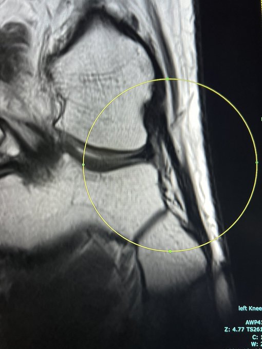
If you see the entire lateral collateral ligament of knee on a single coronal slice, it’s a sign of anterior tibial translation and complete ACL tear.


7
21
171


Ghost sign: MRI sign of osteomyelitis.
It was primarily described to confirm osteomyelitis in diabetic neuropathic arthropathy. However, it can be applied anywhere in bones.
In a non-contrast T1-weighted image, it is described as a poorly defined margin of the bones (Ghost)


2
30
171

