

Fouad Boulos
@fouad_boulos
Followers
2,925
Following
584
Media
313
Statuses
1,660
Professor and Breast Pathology Section Head @wusm_pathology . Trained at Vanderbilt under David Page and Jean Simpson. #breastpath #bstpath
St Louis
Joined April 2019
Don't wanna be here?
Send us removal request.
Explore trending content on Musk Viewer
Arlington
• 738010 Tweets
Elon Musk
• 409634 Tweets
مدريد
• 393862 Tweets
Champions
• 366870 Tweets
Brazil
• 286683 Tweets
#انقاذ_النصر_مطلب_عشاقه
• 269475 Tweets
Colorado
• 242591 Tweets
لاس بالماس
• 190079 Tweets
UEFA
• 168949 Tweets
#UCLdraw
• 168737 Tweets
Beşiktaş
• 125063 Tweets
Xandão
• 84035 Tweets
Bayern
• 84003 Tweets
Bluesky
• 54008 Tweets
#BJKvLUG
• 47787 Tweets
#الاتحاد_التعاون
• 42038 Tweets
McDonalds
• 38715 Tweets
Lugano
• 36390 Tweets
Mustafa Kemal Atatürk
• 28071 Tweets
Las Palmas
• 26072 Tweets
Girona
• 25516 Tweets
Jurassic World
• 21140 Tweets
Rafa Silva
• 19059 Tweets
Brahim
• 16403 Tweets
Servette
• 14710 Tweets
بنزيما
• 14065 Tweets
Semih
• 12755 Tweets


Pinned Tweet

Happy to share the publication of my review article with
@RezaTaha_
regarding the use of E-cadherin in the diagnosis of mammary epithelial proliferations. All feedback welcomed!
@wusm_pathology
@washupathedu
#breastpath
#PathTwitter

4
10
52
@DaniBeckman
Amazing as always. Unfortunately, as Jonathan Swift so eloquently said: Reasoning will never make a man correct an ill opinion, which by reasoning he never acquired.
4
19
247
One in a million Monday. First of all, my apologies to all placental pathologists reading this tweet, this is not personal, your work is noble, and you are appreciated. But I can't say I'm a fan of placental pathology. The reason is the times placental pathology provides any
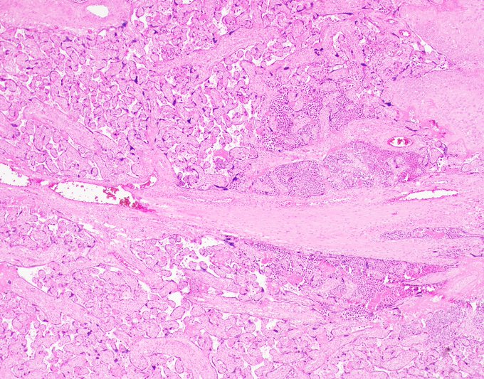
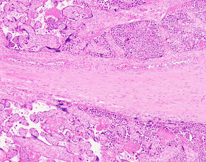
27
79
226
One in a million Monday.
Unwittingly saved by the clinician. This case was not a consult, it was just a good old biopsy from a satellite hospital for whose patients we provide pathology diagnostic services. In that hospital, breast surgeons coordinate breast patient care,
21
80
217
Welcome to my Twitter account where I will be sharing my experience and thoughts from difficult cases received on the WASHU breast consultation service. Questions and comments always welcome. We learn together.
#PathTwitter
#BreastPathology
@wusm_pathology
@washupathedu

13
30
206
Dear pathology community, I am being held hostage by a mob of six unstable juveniles and their leader. I have no access to pathology cases or slides and I’m being coerced into enjoying the Christmas spirit. Please organize a rescue mission promptly. My diagnostic livelihood is on

32
6
186
Dear friends,
Six months ago, I decided to share a few of our consult cases on Twitter in order to promote the consult service and give it an opportunity to grow. The plan was to share one case a week, but as it turned out, discussing interesting cases

11
46
179
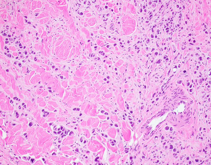
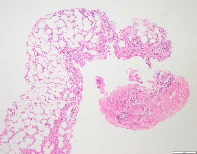
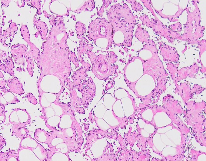
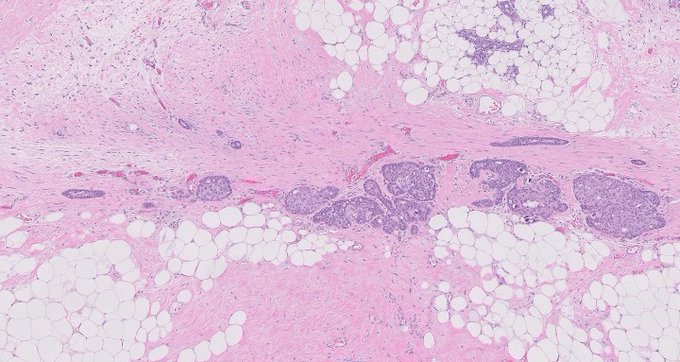
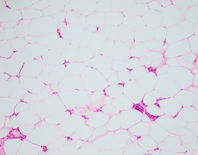
There are two types of pathologists: those who have missed an invasive lobular carcinoma, and those who will. Here's an example of invasive lobular carcinoma masquerading as fat necrosis
@wusm_pathology
@washupathedu
#PathTwitter
#BreastCancer
#BreastPathology
8
71
169
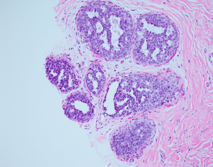
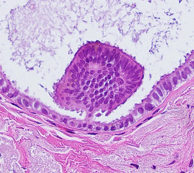
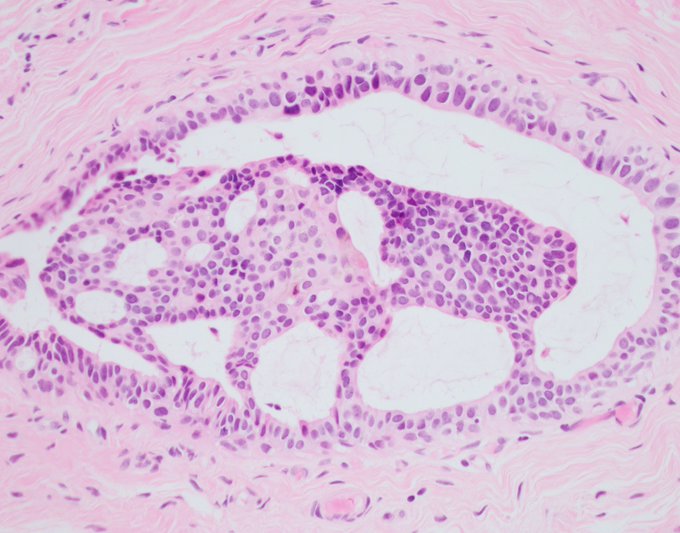
If someone ever asks you for an example of ADH, show them this picture. Uniform round cells, rigid architecture, one space, background columnar cell change, microcalcifications. In other words, perfection.
#breastpath
#PathTwitter
@wusm_pathology
@washupathedu

8
42
161
Today is my birthday and it's International Pathology Day! Somebody tell me this isn't pure destiny :)
#InternationalPathologyDay
61
5
152
Sunday musings. Five things I have learned over the years, cemented by the consult service, that every pathologist should consider when releasing diagnoses into the wilderness.
1. Pathologists are morphologists, yes, but much more importantly, they are consultants. Findings that
8
47
133
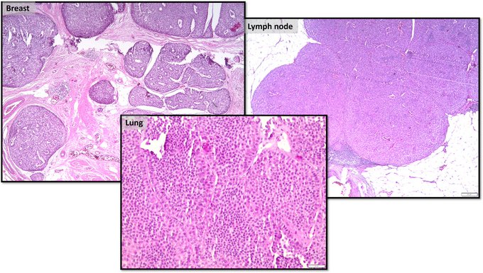
One in a million Monday.
Today we’re bridging two subspecialties
#breastpath
and
#hemepath
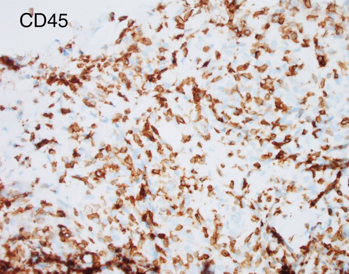
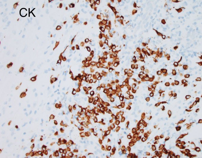
in a rare and unusual case. This is a middle aged patient with known left breast cancer, now presenting with a right sided breast mass and axillary lymph node. Both breast and lymph node
13
45
135
13
13
139
My mentors David Page and Jean Simpson used to tell me this. High-power pathology, low-power pathologist. And this is of course very true. You do not want to get lost in the minutiae because they will steer you off track, most of the time that is. Sometimes though, a closer look
3
39
135
Thank you all for your spot-on responses.
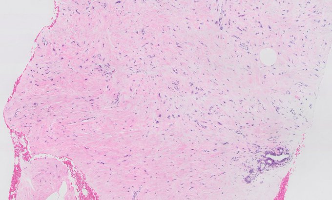
This is indeed a myofibroblastoma in a 75-year-old male patient. This palisaded pattern is quite rare in my experience and in this particular case was very prominent and strongly suggestive of Schwannian tumors. Schwannomas, though they

10
54
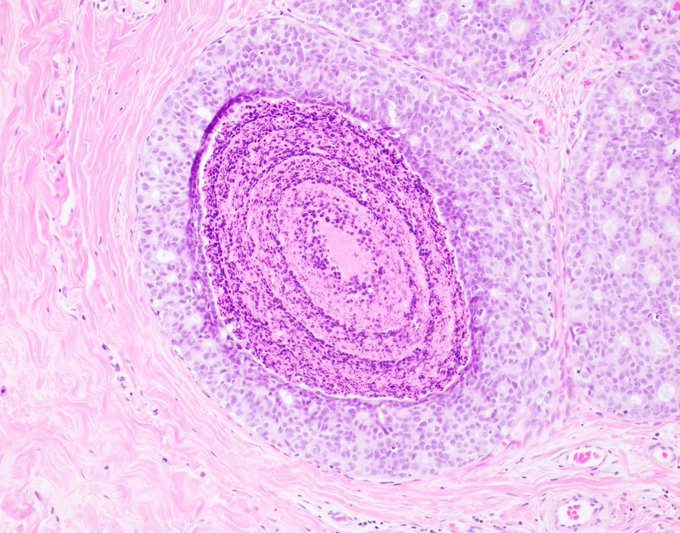
127
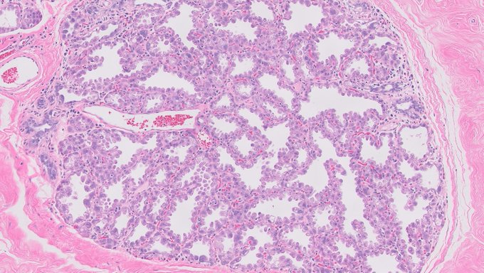
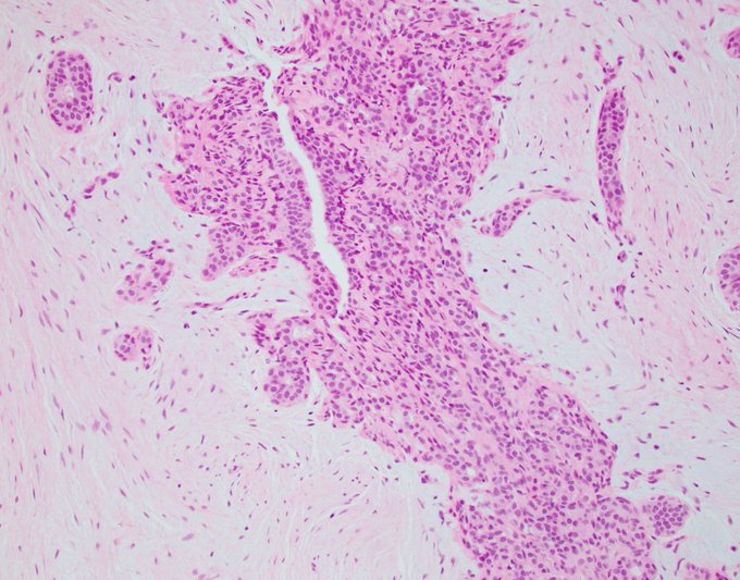
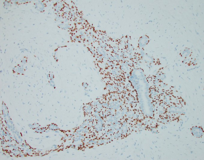
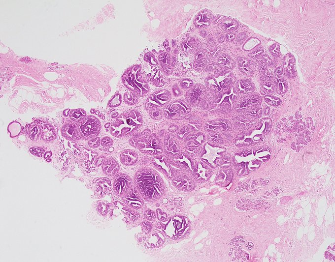
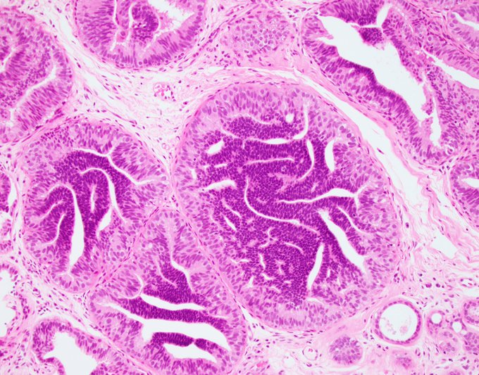
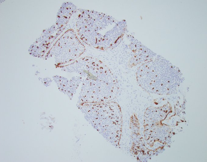
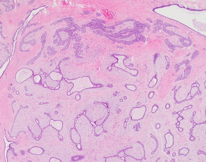
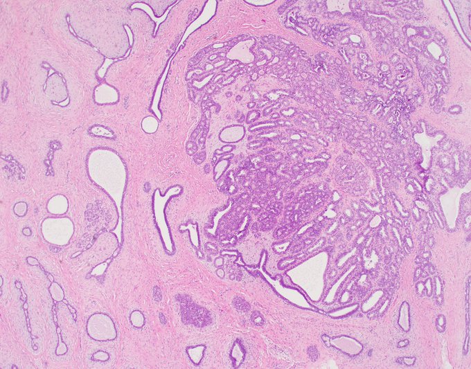
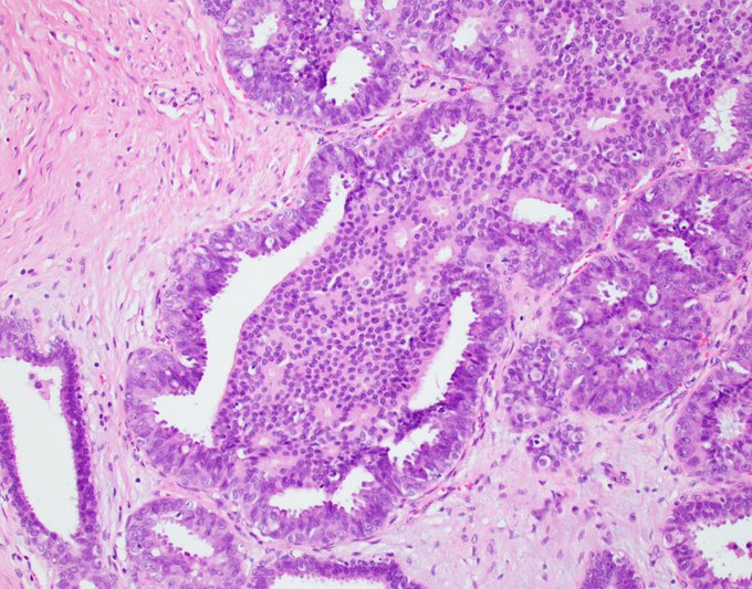
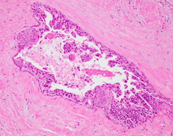
Papillary lesions are a hairy affair! How would you characterize this papillary proliferation with preserved myoepithelial cells? No poll this time, and I am not quizzing, I am asking for help :)
@wusm_pathology
@washupathedu
#PathTwitter
#breastpath

19
47
124
Going back to the UDH/DCIS differential may feel like beating a dead horse. But the cases keep coming and the implications are considerable. Yesterday's tweet illustrated a case diagnosed as DCIS by a skilled and experienced pathologist who was made acutely aware of his mistake
9
47
126
One in a million Monday.
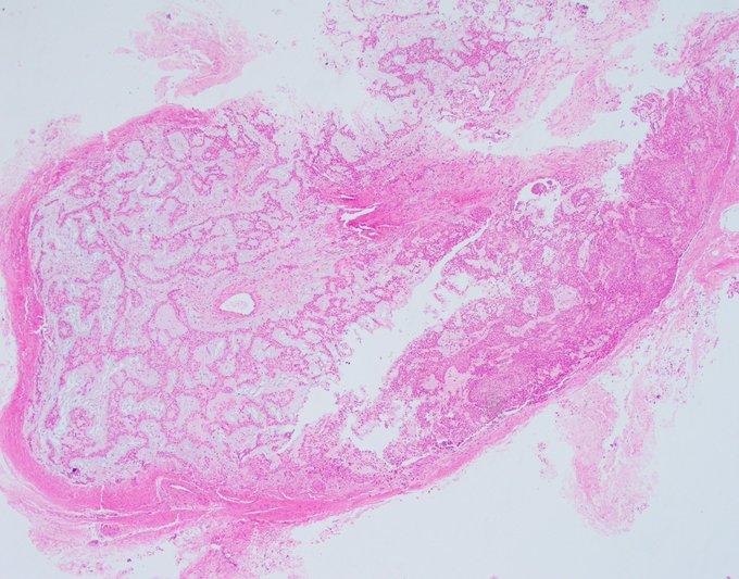
This case stayed on my desk and made the rounds multiple times for several days because it is unlike any case I have ever seen. Cystic hypersecretory spaces, TDLU-like architecture, relatively bland cytology, this lesion was screaming benign off the top
11
41
124
The most well-differentiated invasive carcinoma that ever lived.
Node the background of flat epithelial atypia and atypical ductal hyperplasia. There is atypical lobular hyperplasia elsewhere as well. Classic Rosen triad.
@wusm_pathology
@washupathedu
#BreastCancer
#breastpath

4
34
124
Don't cross these flimsy Roman bridges toward a diagnosis of atypical hyperplasia. They will crumble under your feet. UDH mimicking ADH. Key findings: streaming overlapping variable nuclei and non-rigid arches
#BreastPath
#PathTwitter
@wusm_pathology
@washupathedu
3
36
121
Another ADH vs. DCIS borderline case.
Stay tuned for my thoughts on such cases tomorrow!
@wusm_pathology
@washupathedu
#breastpath
#PathTwitter
#PathX
#pathology

17
26
116
Thank you all for your comments. As I expected, the responses were mixed, but ADH was a clear winner in the first case, in part driven by the need and recommendations to be more conservative in our core biopsy diagnoses, and in part because of the presence of two cell

13
35
117
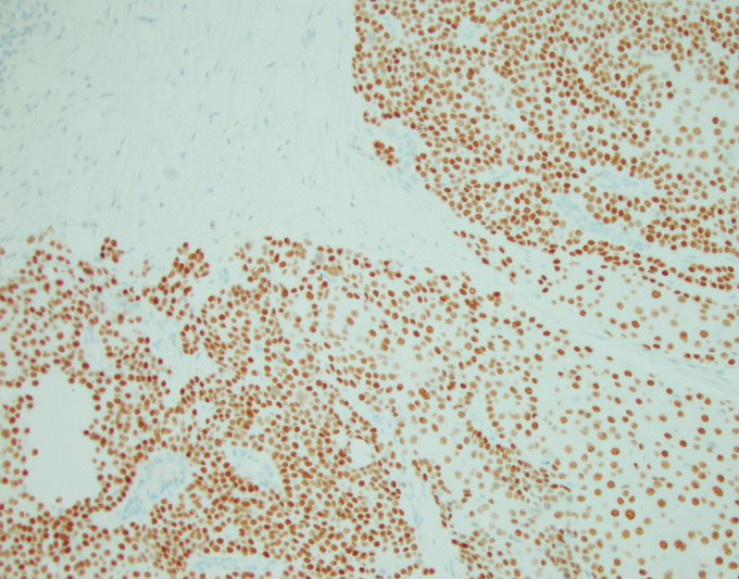
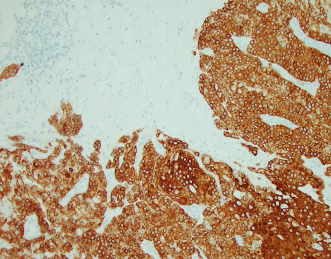
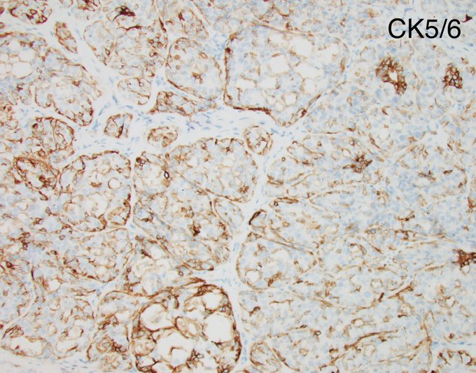
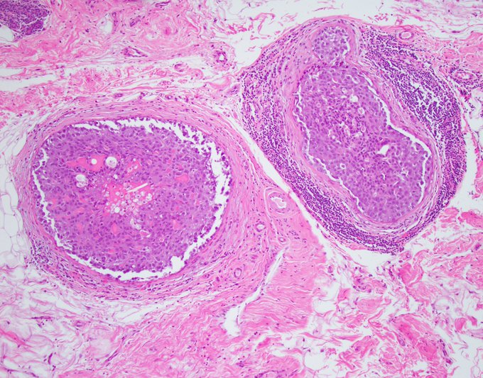
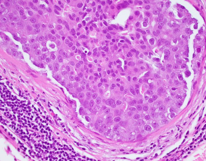
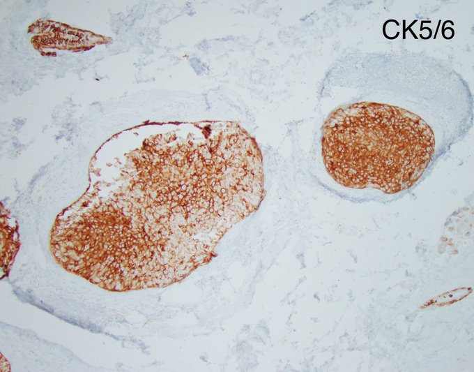
This is a consult case that was already stained. The question was: Could this be UDH given the mosaic-pattern of CK5/6 positivity (image 2)?
The answer is NO, because the cytology is malignant. Basal-like DCIS can express CK5/6 as seen in this case, either diffusely or variably.


8
30
116
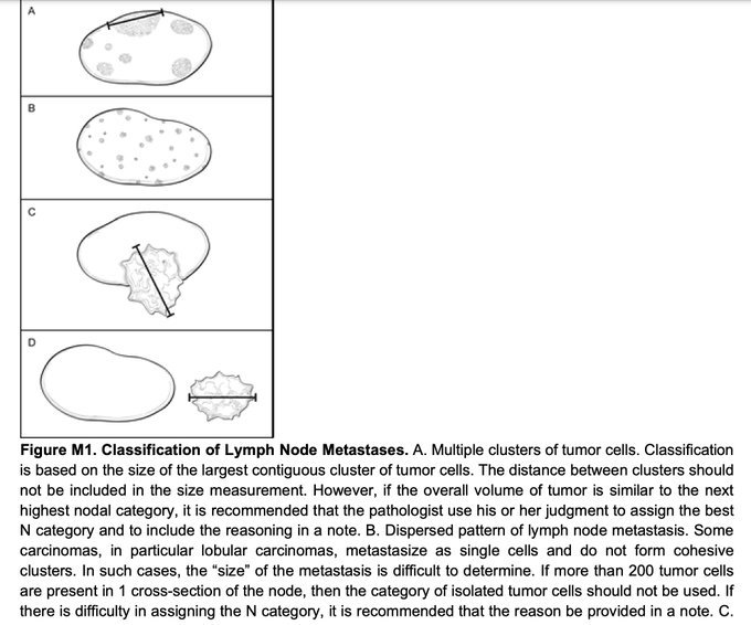
Friday staging mini-Tweetorial!
Staging in breast cancer is a subject dear to my heart that I feel is often confusing and misunderstood. The reason is simple. On a slide, we see things in two dimensions, and therefore we are taught in two dimensions and we come to remember and
7
50
116
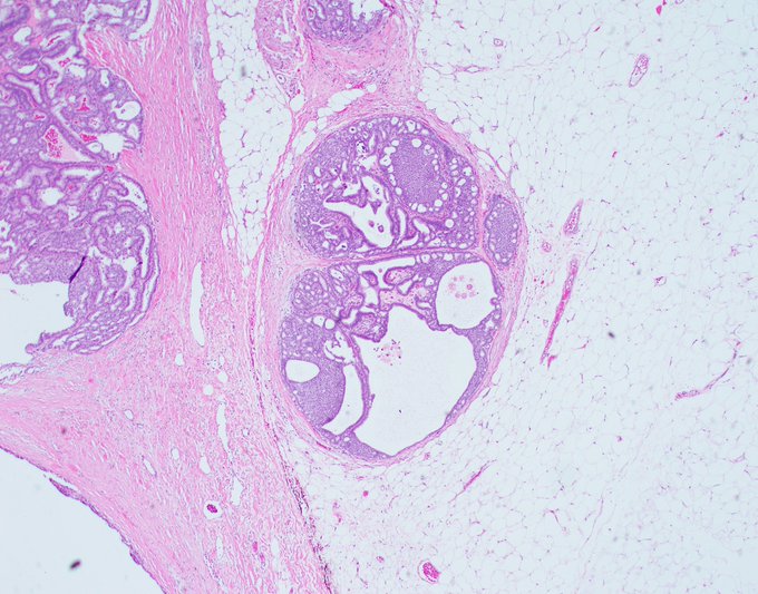
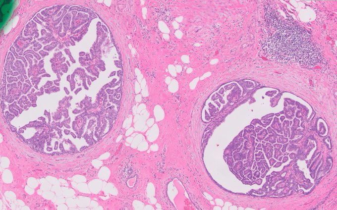
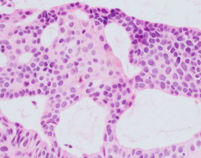
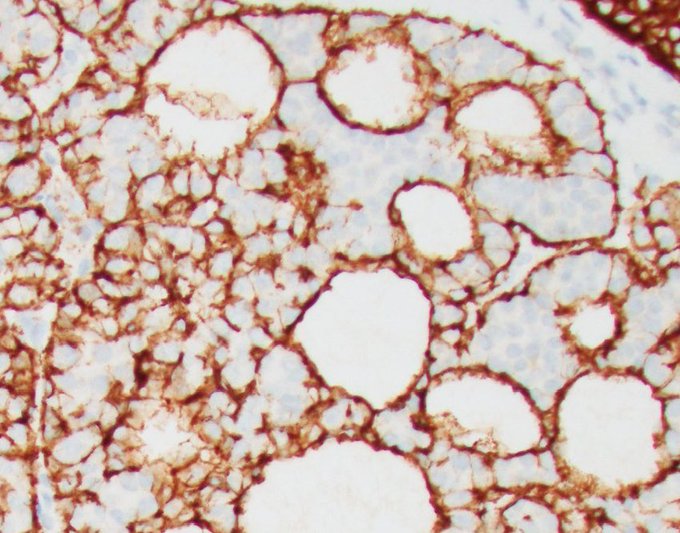
My story with solid papillary carcinoma (SPC) is marginally interesting, so I will go ahead and share it with you innocent onlooking victims 🙂
SPC was first described by Dr. Horacio Maluf in an AJSP paper in 1995 (which I shall go back to towards the end of this tweet). Yet,
19
26
113
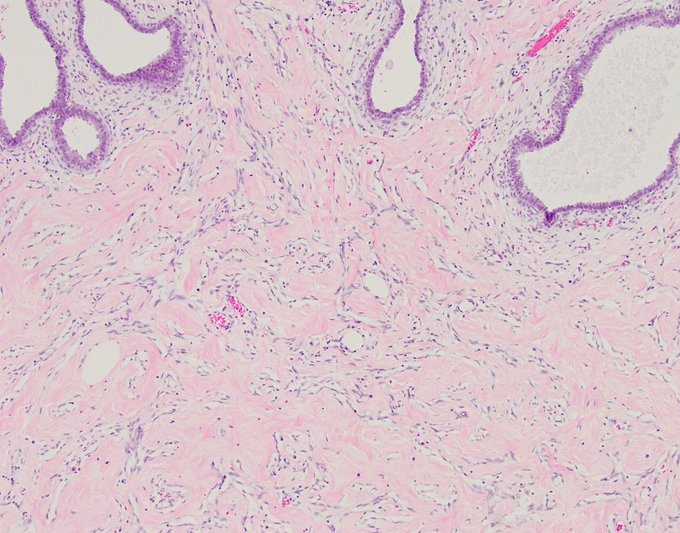
Let’s discuss the PASH vs. Fibroadenoma post.
PASH is a very popular entity. Just like stromal fibrosis, residents and attendings love to throw it in the mix of benign diagnoses in a biopsy report.
But I’m not a fan.
To be fair, it does have a clever name, a catchy acronym,

11
38
113
Friday mini-Tweetorial! Part 2
Question: Is the absence of myoepithelial staining equivalent to invasion?
Answer: No.
Explanation: We cannot equate a lack of myoepithelial staining with invasion, first because lack of staining does not mean a lack of myoepithelial cells,
4
38
112
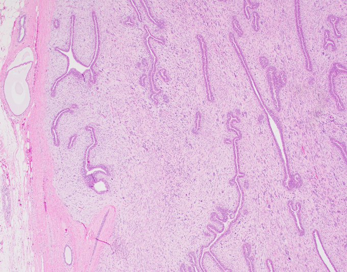
Beautiful example of periductal stromal condensation in a phyllodes tumor
#breastpath
#PathTwitter
@wusm_pathology
@washupathedu

1
28
110
Adenomyoepitheliomas are rare, challenging, biphasic neoplasms with a real potential for atypical change and malignant transformation. My mentor, David Page, would beg to differ because he was the most benign pathologist to ever walk the face of the earth. In my experience, about
1
32
106
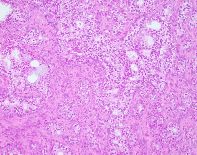
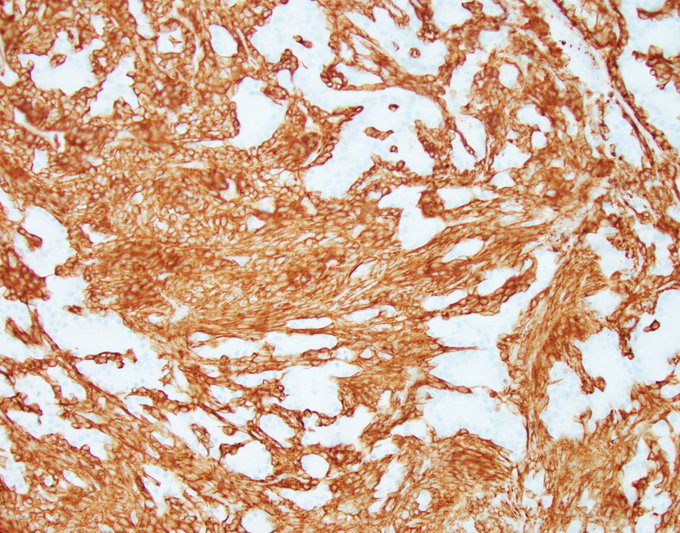
Easing into Monday morning with an eye-catching histologic pattern, uncommonly seen in this mammary neoplasm. What is your differential diagnosis? What IHC panel would you like to perform?
@wusm_pathology
#PathTwitter
#Breastpath
16
40
104

20
16
103
Thank you all for your participation and for helping me demonstrate a very helpful finding when it comes to differentiating invasive vs. non-invasive disease.
The image depicts invasive carcinoma with DCIS-like morphology circling and impinging on a normal duct. The distribution
8
33
101
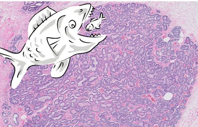
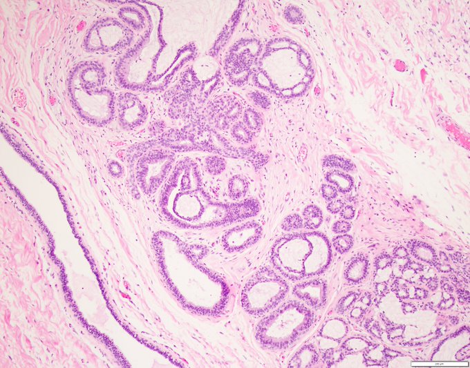
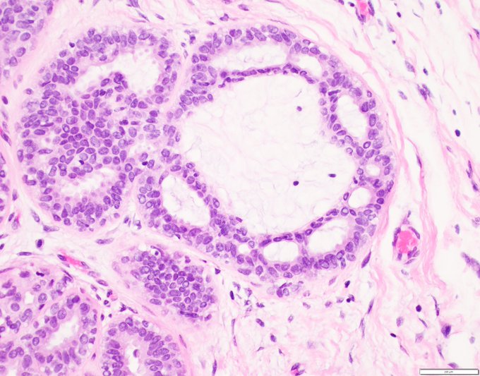
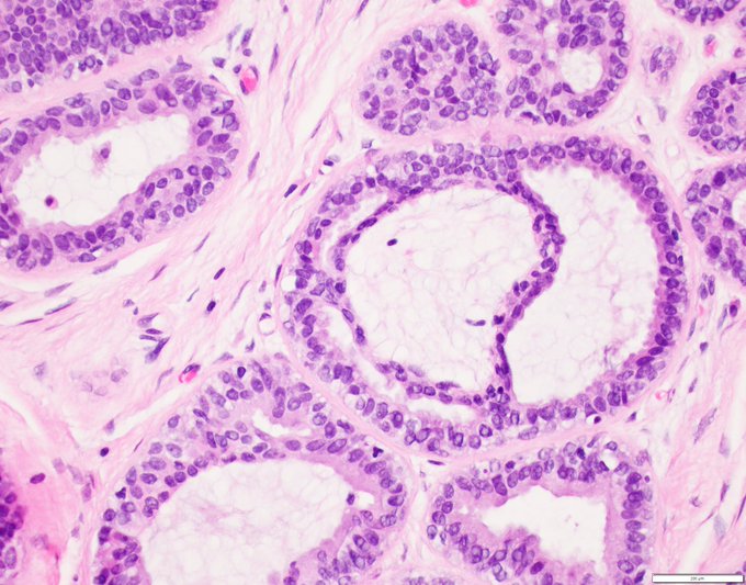
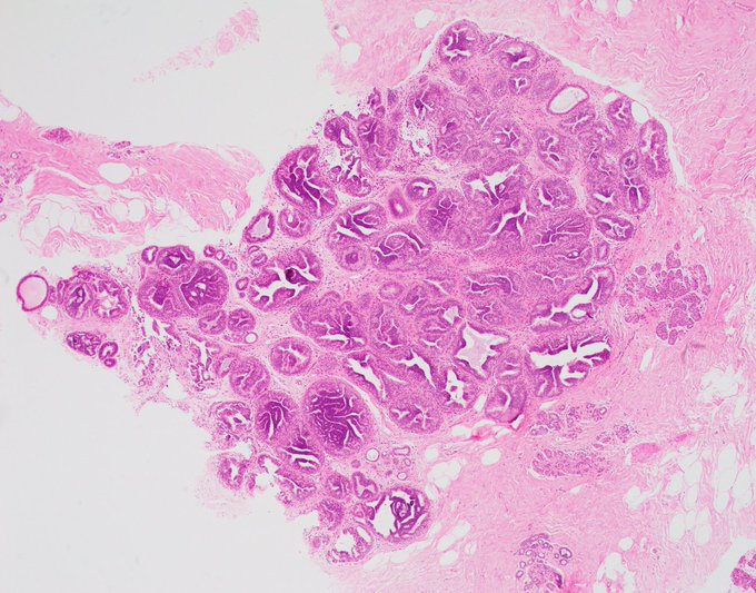
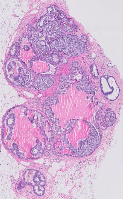
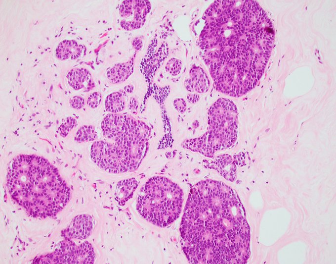
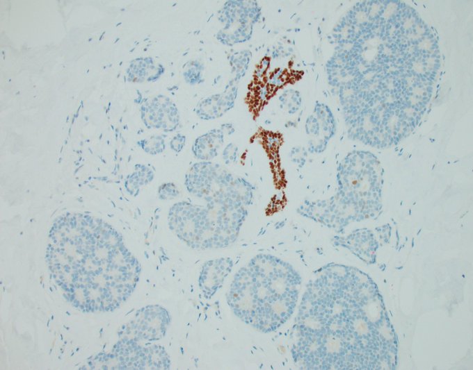
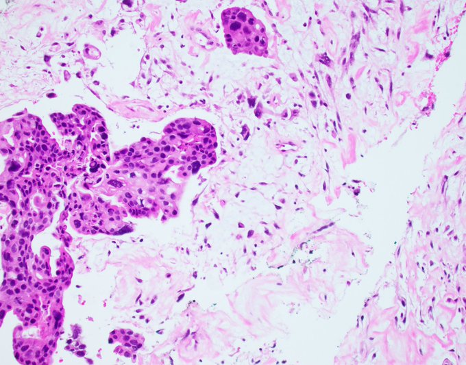
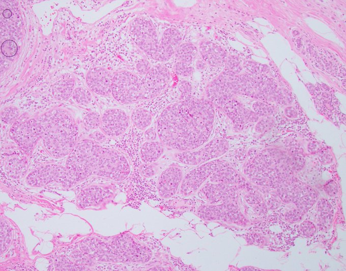
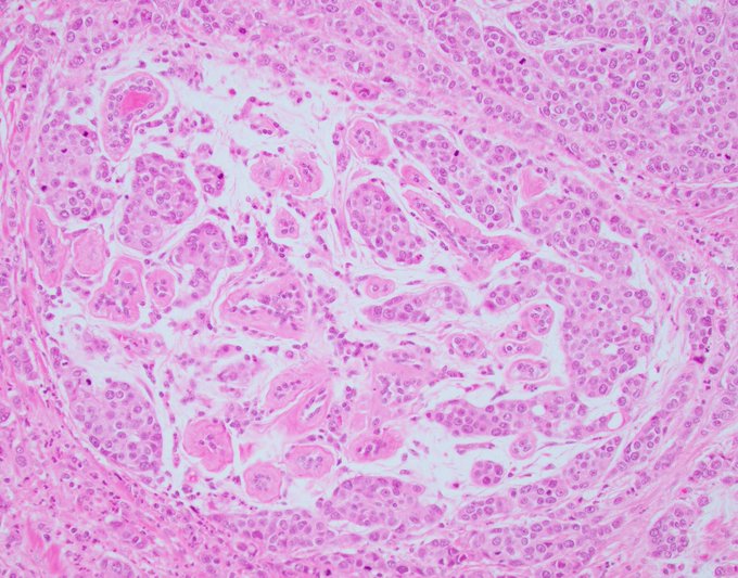
Benign morphology, almost benign biology in the breast, often deadly biology in the salivary gland.
The conundrums of neoplasia will never be resolved.
#breastpath
#PathTwitter
@wusm_pathology
6
22
102
Peripheral slit-like spaces and a glomeruloid architecture are indicative of usual ductal hyperplasia, until they're not.
Here's some glomeruloid intermediate-grade ductal carcinoma in situ for your eyes to feast on.
#PathTwitter
#breastpath
@wusm_pathology
@washupathedu
2
26
102
Single focus in a single core from the breast of a 45-year-old woman.
Thoughts?
@wusm_pathology
@washupathedu
#breastpath
#PathTwitter
#PathX
30
21
100
25
18
98
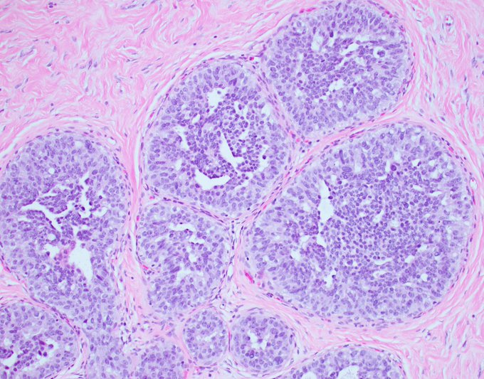
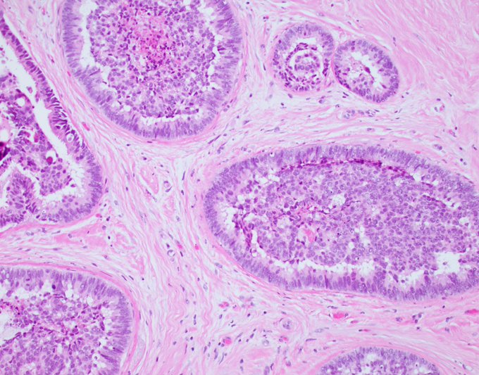
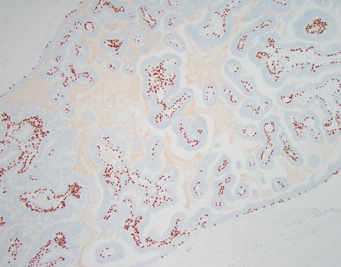
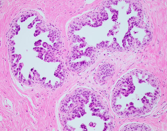
Thank you all for your input. The problem with a diagnosis of papillary DCIS is the diffuse presence of myoepithelial cells in the fibrovascular cores, which should not happen in a neoplastic papillary proliferation. In this case, I elected to still go for DCIS based on the

5
20
98
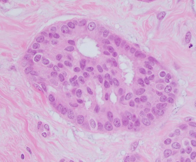
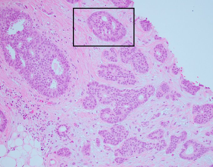
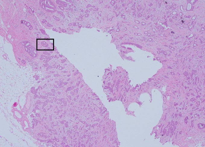
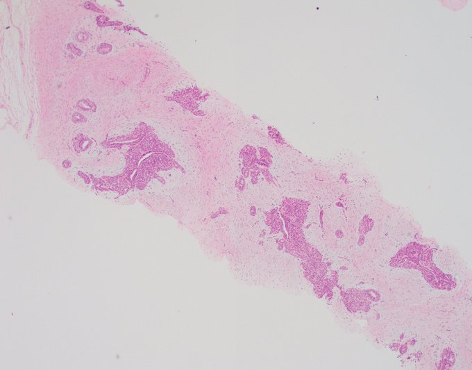
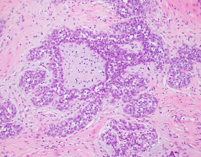
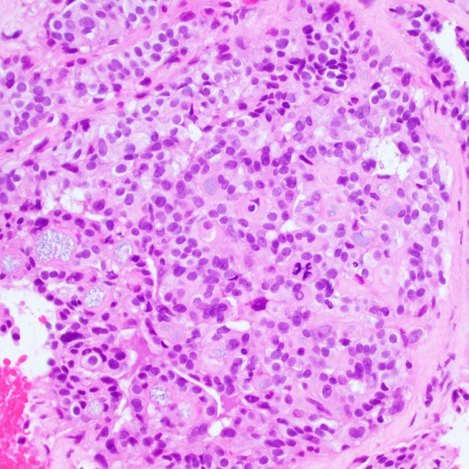
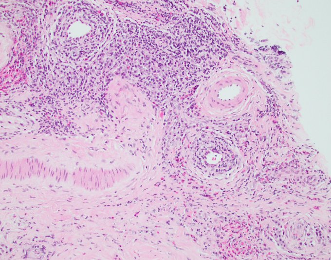
I heard late Dr. Juan Rosai during a course he gave in Italy joke about the need to combine breast and prostate pathology once and for all. It was funny, and of course, partly true. The attached images are from a biopsy that shows literally five atypical glands lacking
1
20
97
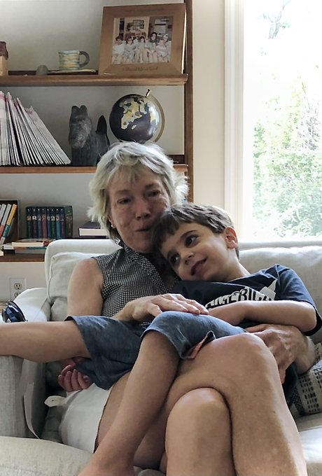
Gratitude Sunday.
This is Dr. Jean Simpson with my son Karl. She was in St. Louis for the weekend and stopped by to say hi.
Jean is not only the reason I am tweeting this, but the reason this Twitter account exists. She’s also the reason I am a breast pathologist.
She and
9
4
99
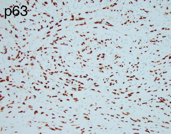
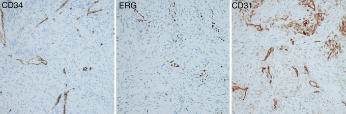
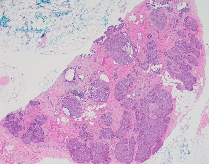
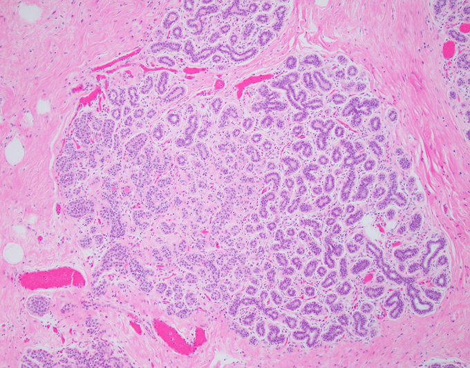
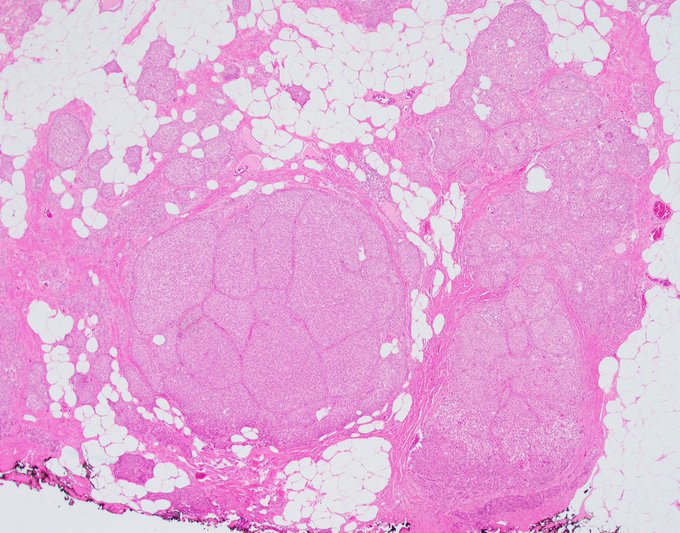
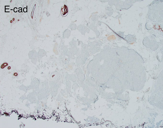
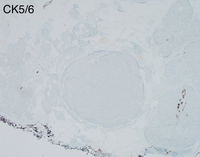
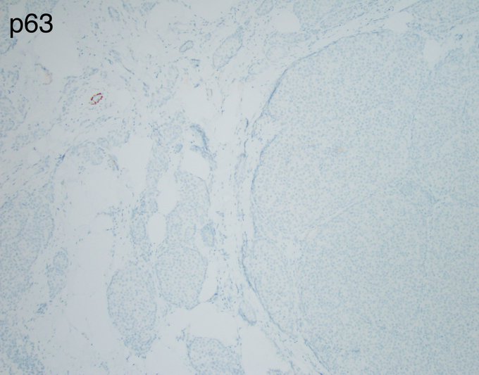
Excision of a breast mass.
IHC = CK5/6 and p63.
What is your diagnosis?
@wusm_pathology
@washupathedu
#breastpath
#PathTwitter
#PathX
25
35
97
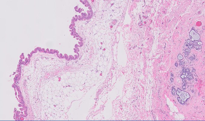
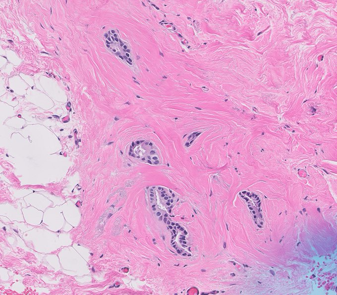
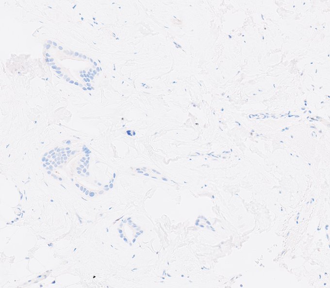
Happy Labor Day to the most industrious organ, producer of the elixir of life, the reason we exist as a species, the breast.
- Lactational change in a breastfeeding woman.
4
13
94
Long weekend over, time for another game, yay 🙂. What am I trying to demonstrate with this image?
@wusm_pathology
@washupathedu
#PathTwitter
#breastpath
#pathology

29
24
94
Really peculiar pattern of a rare neoplasm.
What is your diagnosis?
#breastcancer
#breastpath
#PathTwitter
@wusm_pathology
@washupathedu


13
26
93
We've seen it a few times, but it's always fun to see. And it will always make us pause. Usual ductal hyperplasia with central necrosis.
@wusm_pathology
@washupathedu
#breastpath
#PathTwitter

3
23
93
Tuesday’s quiz was not fair, because it was not intended to get a correct answer but to illustrate two things. First, the importance of context, second the limitations of morphology.
1. Context is everything. A lion at the zoo is not a lion in the living room, a swimsuit on the

8
25
93
Photogenic Friday.
Fibroadenoma with periductal adenosis and myoepithelial hyperplasia.
Behold the fibroadenomyoepithelioma!
🙂
@wusm_pathology
@washupathedu
#breastpath
#PathTwitter
#pathology
5
28
93
I have been avoiding this topic for a while, but I think it’s time to ease into it. This may take a couple or more tweets, it certainly deserves a few. So let’s get right into it with this pictorial introduction.
ADH or DCIS? and very importantly, why?
Talk to you soon!
22
21
93
Thank you all for participating in yesterday’s case.
It is indeed a complex fibroadenoma with both atypical ductal hyperplasia (I did not pull the trigger on DCIS) and atypical lobular hyperplasia. This is a very rare combination although atypia in a fibroadenoma is common

13
26
92
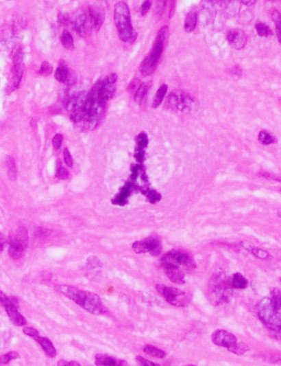
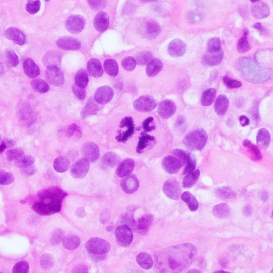
Context is everything.
One of the stories I often tell my trainees is that, as an intern, I saw a tripolar mitosis, the most perfect I had and have ever seen, right at the edge of... a gouty tophus. I was so excited I shared it with my attendings and got a good laugh out of
4
31
90
17
18
89
Thank you all for playing along.
This is indeed usual ductal hyperplasia involving a complex sclerosing lesion. This case illustrates how benign processes can be alarming both radiologically (spiculated mass) and histologically (irregular nests with compact florid epithelial


4
22
90
Do not mistake peripheral palisading in invasive carcinoma for a preserved myoepithelial layer.
Invasive solid papillary carcinoma with peripheral palisading (pseudo-myoepithelial layer).
@wusm_pathology
@washupathedu
#breastpath
#PathTwitter
#PathX

1
19
88
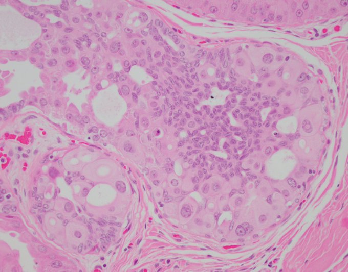
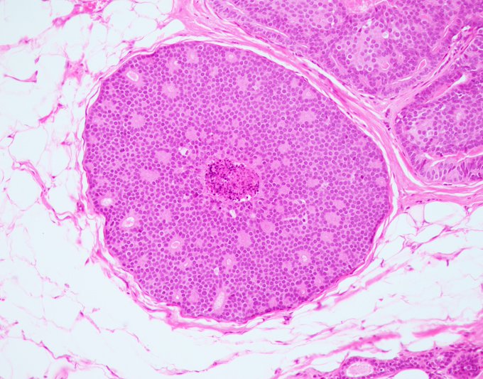
Beautiful Low-grade DCIS with rosettes and central necrosis.
1. Please don't call this comedonecrosis, it may imply a higher-grade more aggressive process.
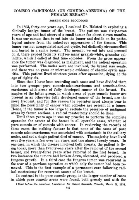
2. Remember that comedo carcinoma, coined by Dr. Bloodgood in 1934, refers to a gross finding in mastectomies when
3
29
87
Cytokeratin can be picked up by mast cells in a sentinel lymph node, potentially leading to a false-positive diagnosis, which is what happened in this case.
Clues to the correct diagnosis include:
1. Subtle but distinct granularity of the cytoplasm
2. Lack of membranous

2
21
88
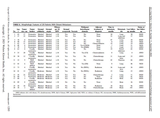
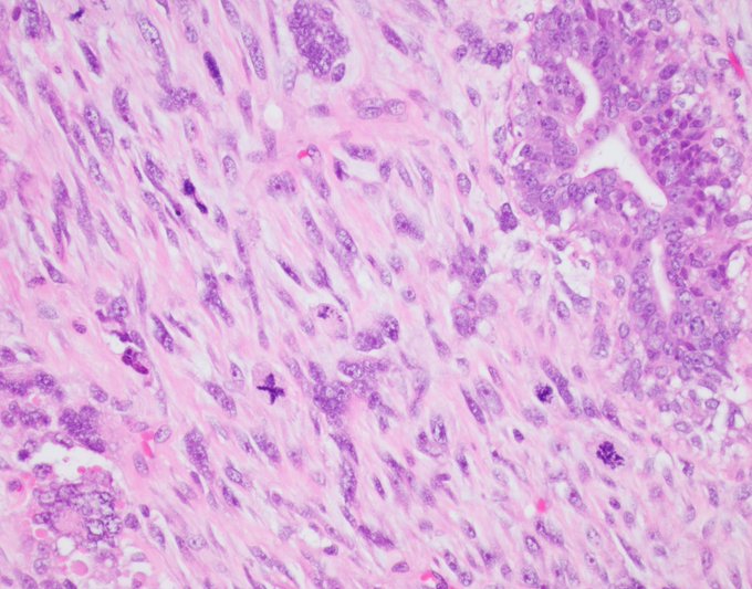
It's a minefield out there. Imagine having this on a biopsy. Malignant phyllodes tumor with a single cell epithelioid infiltrative pattern mimicking carcinoma/EHE/MM... It's a miracle we still dare to practice
#PathTwitter
#BreastPath
#breastcancer
@wusm_pathology
@washupathedu
5
24
87
Metaplastic carcinoma of the breast can take on a variety of different morphologies, and I have seen pseudovascular patterns with the acantholytic variant of squamous cell carcinoma, but I have never seen the epithelial/glandular component of a metaplastic carcinoma adopt a
7
27
87
No IHC, no HP. Only this low-power view.
What say you?
@wusm_pathology
@washupathedu
#breastpath
#PathTwitter
#PathX
35
22
87
How do you recognize atypical lobular hyperplasia on low power?
#breastpath
@washupathedu
#PathTwitter
9
17
86
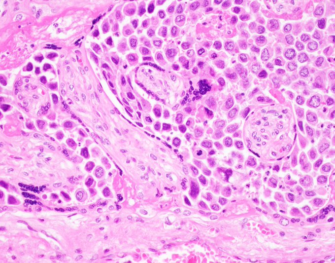
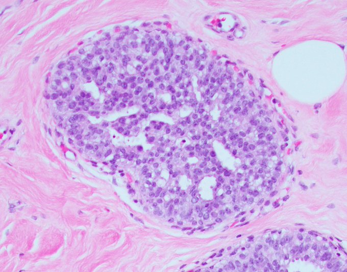
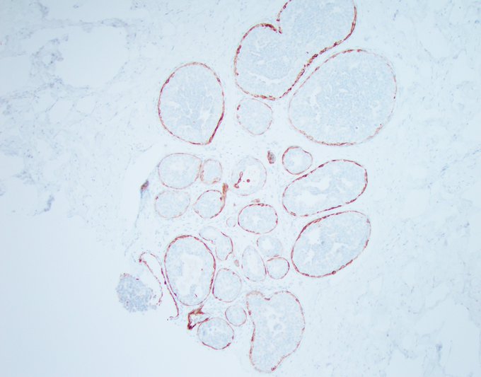
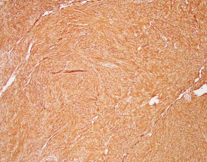
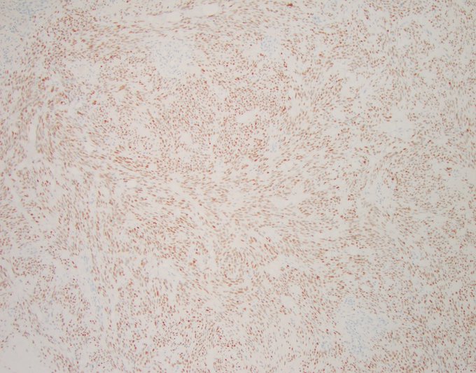
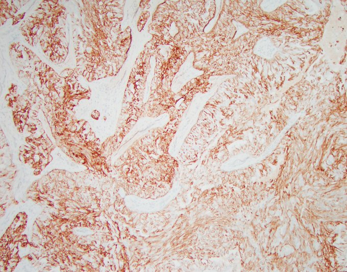
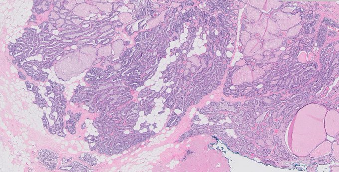
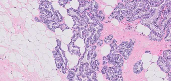
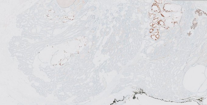
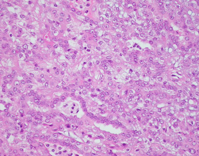
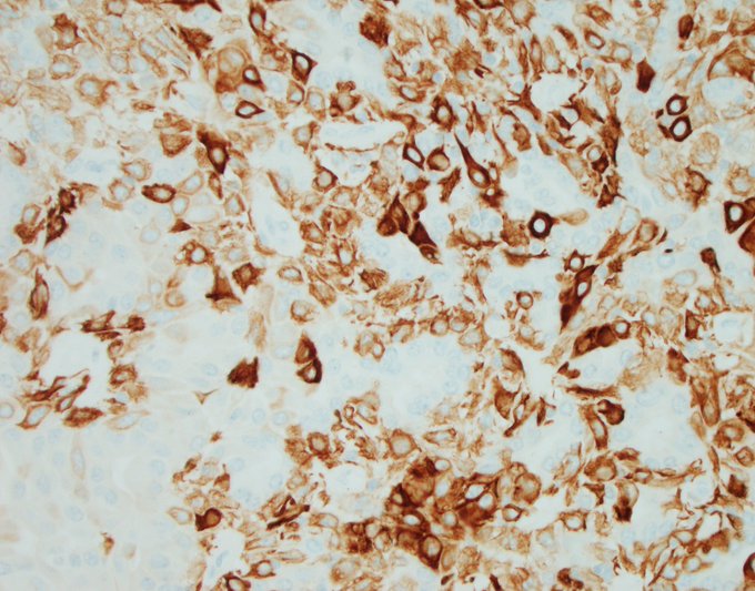
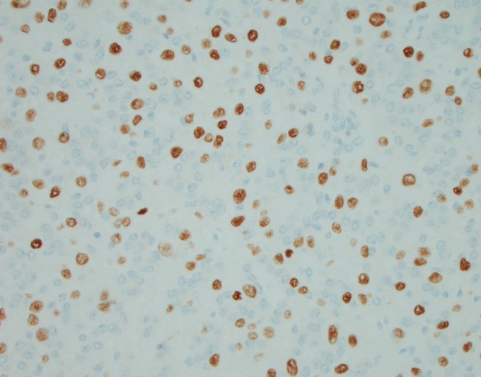
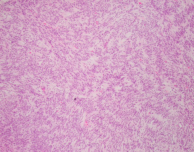
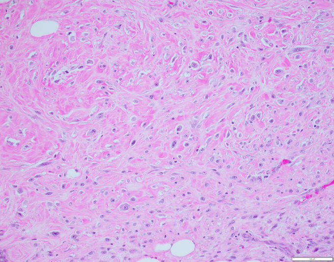
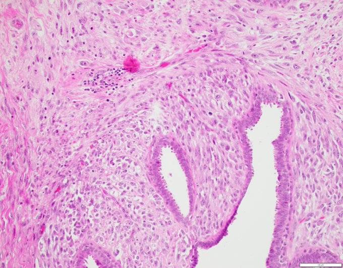
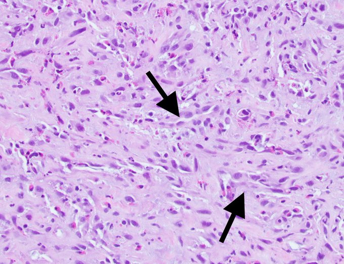
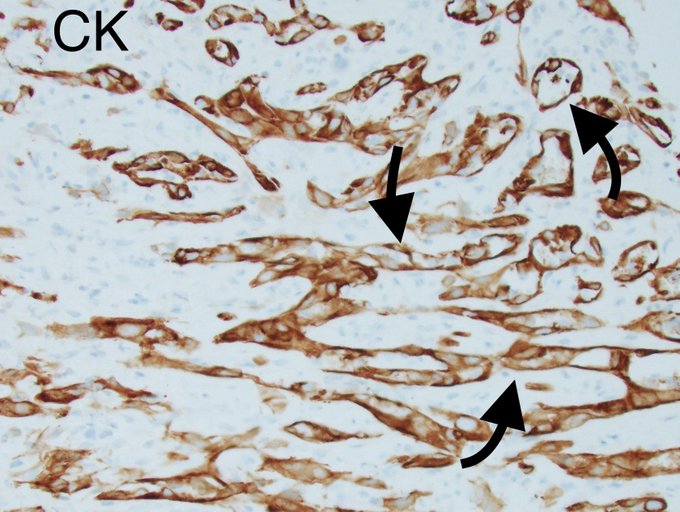
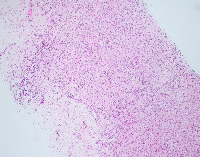
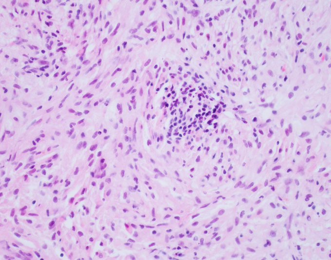
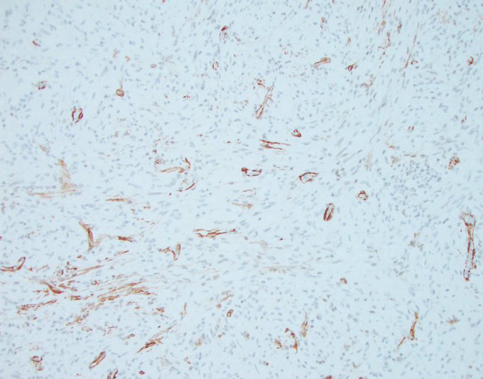
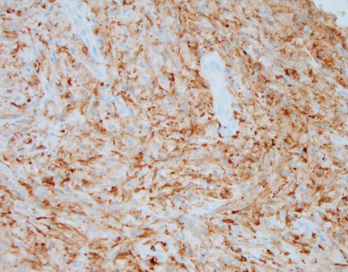
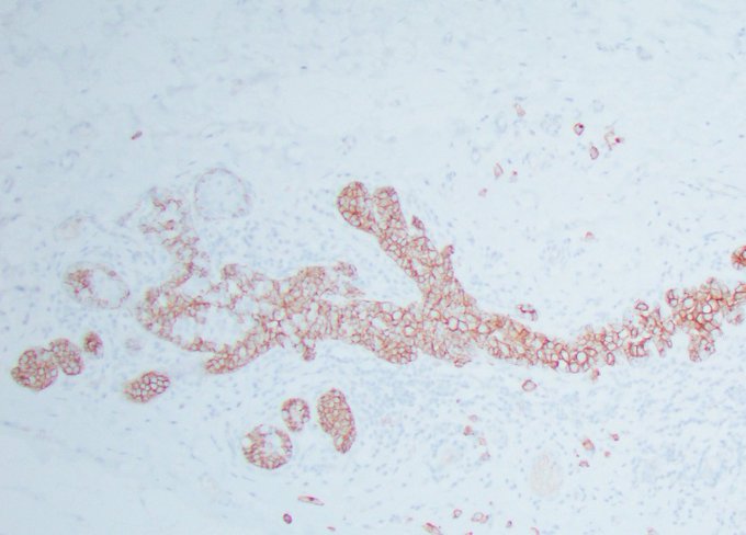
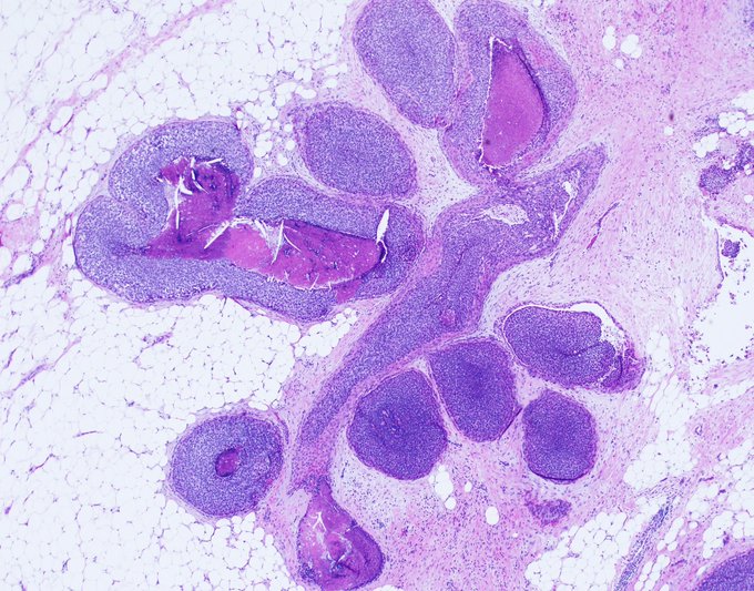
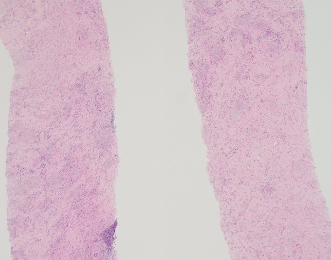
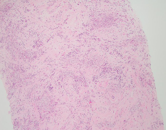
Morphologic Monday
This consult case is on an older woman with a palpable breast mass. Appropriately, the differential diagnosis of the consulting pathologist is metaplastic carcinoma, malignant phyllodes tumor, and primary sarcoma.
Immunohistochemistry is non contributory.
15
23
85
Friday Mini-Tweetorial: Part Deux continued
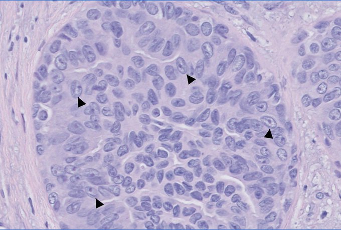
3. Reptilian heads, or dinosaur heads if you're like my son, which refers to UDH nuclei with a central long groove and an eccentric nucleolus in lieu of the mouth and eye. I used a similar analogy for papillary thyroid carcinoma, and
7
20
85
Biopsy of a breast mass in a middle-aged woman.
IHC = CK5/6.
What is your diagnosis?
@wusm_pathology
@washupathedu
#breastpath
#PathTwitter
#PathX
25
19
86
Happy Tuesday PathTwitterverse!
What is your diagnosis and what is its significance?
@wusm_pathology
@washupathedu
#PathTwitter
#pathology
#breastpath

19
24
85
This is a video briefly explaining my interpretation of the previous post.
Don't forget the popcorn! :)
@washupathedu
@wusm_pathology
#breastpath
#PathX
#PathTwitter
13
16
83
Friday Xposé 🤓
We’re all familiar with the difficulty in properly classifying fibroepithelial lesions of the breast.
Distinguishing fibroadenoma (FA) from benign phyllodes tumor (PT) is what we struggle with most often, and that’s because most of us accept the idea of a
8
27
83
Thank you all, as always, for your comments and interactions. What a lonely place Twitter/X would have been if we were to share cases and get crickets.
This biopsy came to me with a preliminary diagnosis of nodular fasciitis, and I wasn't opposed to that interpretation, but
9
15
83
Thank you all for voting. Briefly, half of 230 participants diagnosed this focus as ADH, a quarter diagnosed it as UDH with apocrine change, and the remainder were split.
The issue with this case (I do not claim to know the final answer) is that I cannot identify a clonal
9
17
82
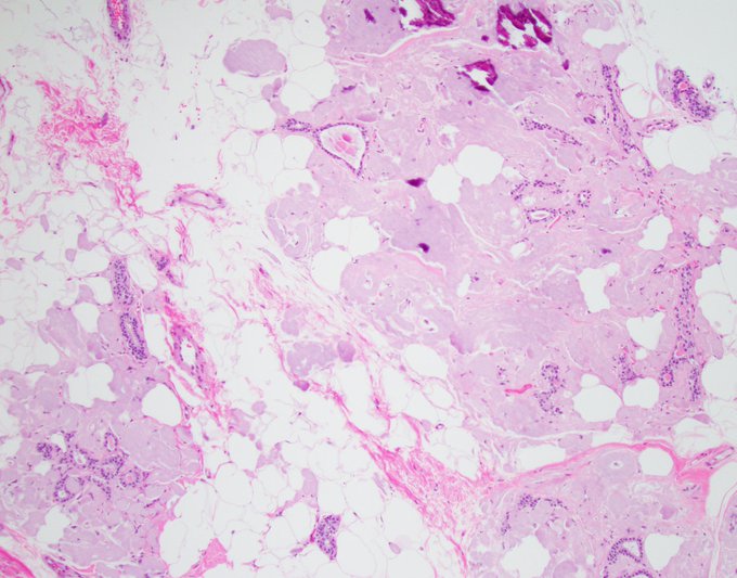
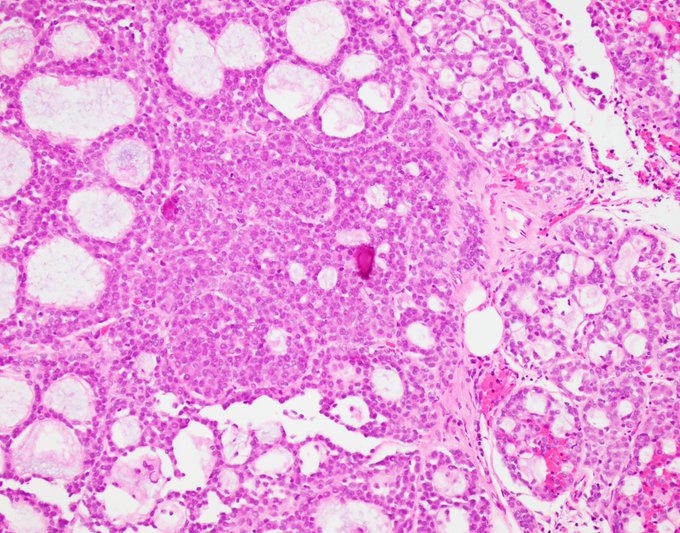
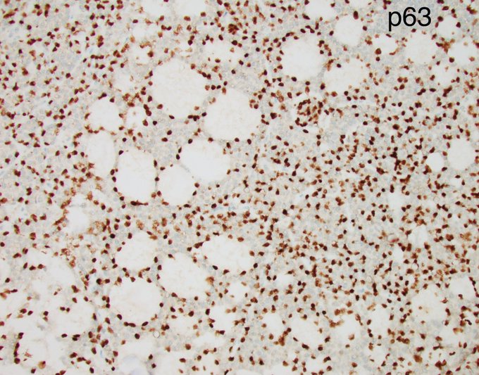
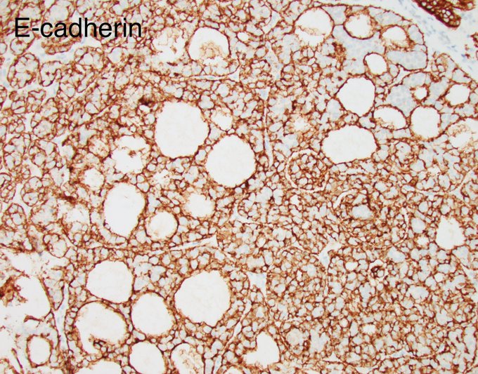
The diagnosis for yesterday’s case is LCIS with collagenous spherulosis. This case is particularly rich in myoepithelial cells, hence the many p63 positive nuclei and the excessive E-cadherin positivity obscuring the negative lobular cells. This amplifies the pitfall of calling
5
16
82
When my 3-year-old daughter was throwing a tantrum yesterday at a breakfast place in Chicago, I tried, for once, to be a better, calmer parent.
I carried her outside to beautiful weather and stood by a small park where pigeons were picking their breakfast in the grass under a
6
3
82
Photogenic Friday.
Apple tree on a misty day 🙂
@washupathedu
@wusm_pathology
#breastpath
#pathtwitter
2
13
80
Papillary lesions are often challenging, and it's my general conviction that they are poorly understood and poorly defined. Sometimes though, you come across a case that is rare and challenging, but can at least be diagnosed with confidence. There's nothing quite like these

3
22
80
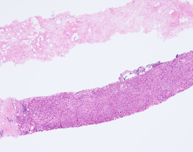
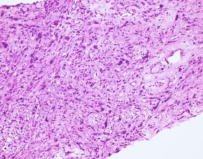
Not all that glitters is gold! This is a biopsy from a 2 cm mass that was called metaplastic carcinoma by yours truly based on histologic pattern and positive p63 and cytokeratin. Excision followed and low and behold...
4
36
80
What is your low power diagnosis of this breast lesion?
#breastpath
#PathTwitter
#PathX
@wusm_pathology
@washupathedu
29
18
79
Sunday reflections.
When I interview applicants for medical school, I ask them what is the most important quality for a physician to have. The most common response I get is empathy, which is a good answer. But the answer I look for but I never get is humility.
Five reasons
5
16
77
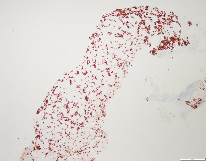
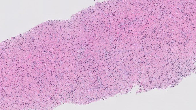
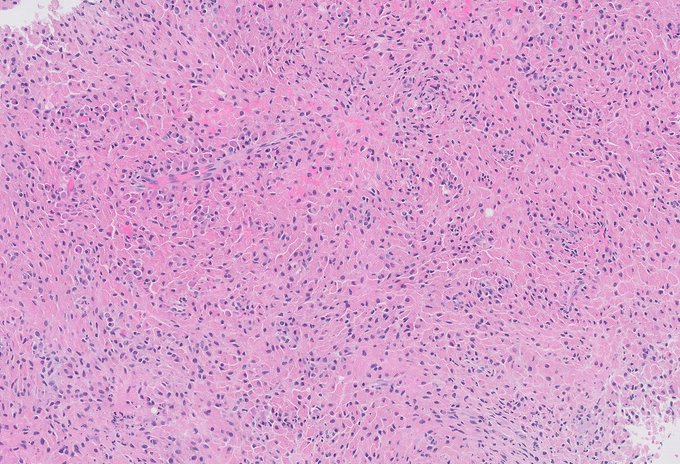
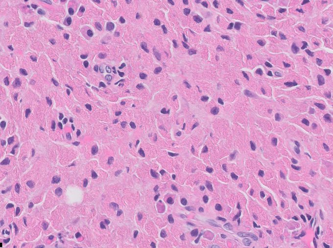
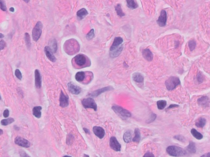
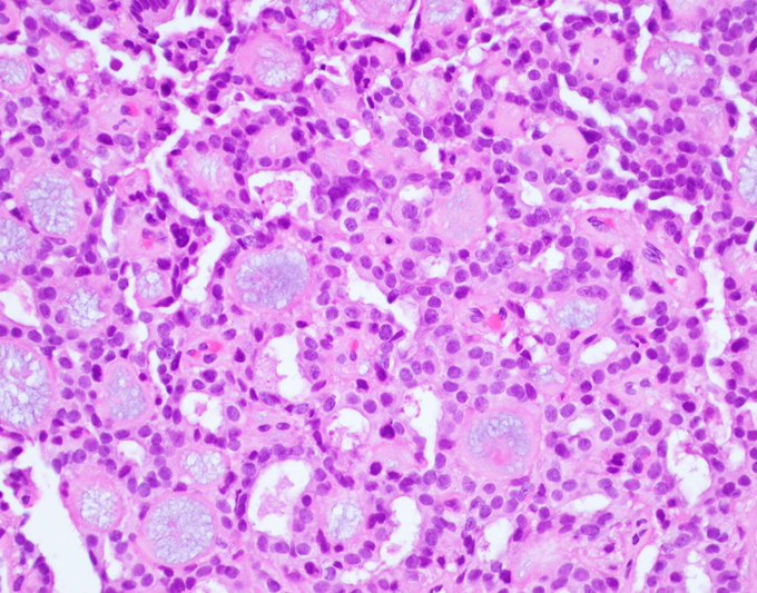
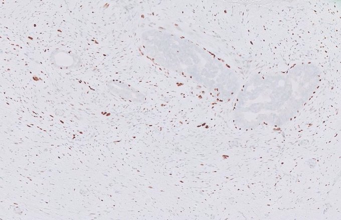
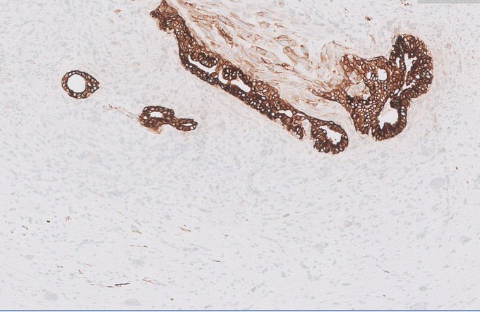
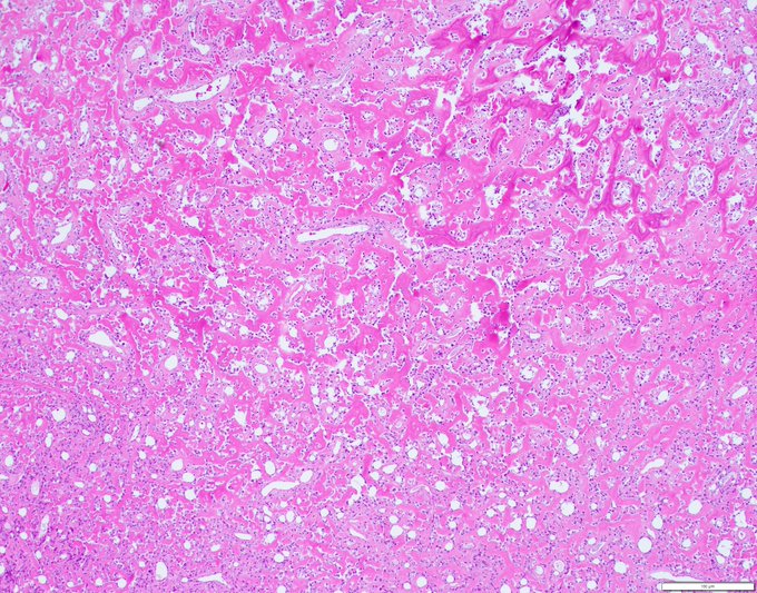
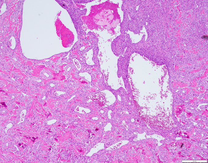
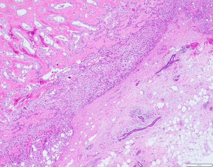
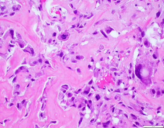
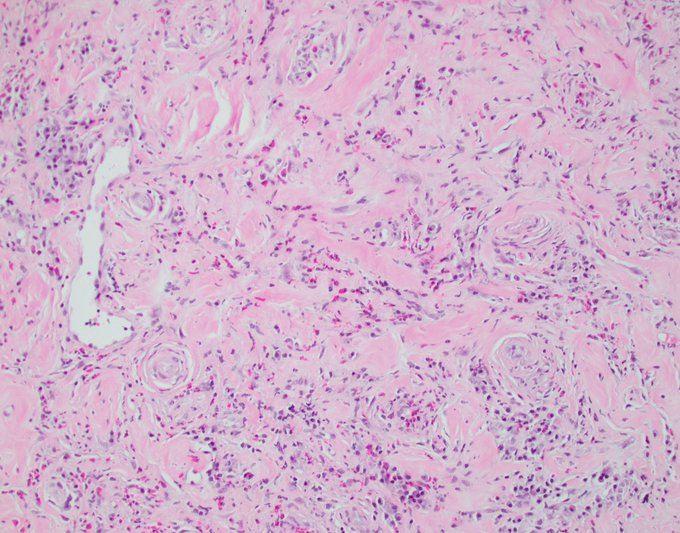
Thank you all for the outstanding responses! This is indeed crystal storing histiocytosis (CSH) with a main differential of granular cell tumor. Exceedingly rare in the breast, CSH can affect any organ and is usually associated with lymphoproliferative disorders and plasmacytic

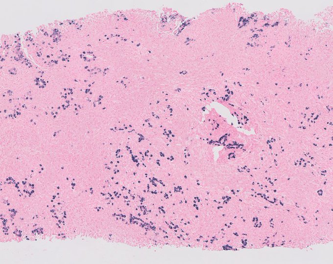
2
18
76
The case I posted yesterday is a resection on a biopsy that was called LCIS.
The key in this case of lobular neoplasia is to remember that E-cadherin, though often weaker than in epithelial cells, is usually retained in myoepithelial cells and its complete absence should make
3
21
76
50 year old woman with left breast microcalcifications.
Diagnosis?
@wusm_pathology
@washupathedu
#breastpath
#PathTwitter
#PathX
14
17
76
Breast biopsy for calcifications. What is your diagnosis?
@wusm_pathology
@washupathedu
#breastpath
#PathTwitter
#PathX
22
20
76
Single focus on core biopsy.
What is your diagnosis? See poll below
@wusm_pathology
@washupathedu
#breastpath
#PathTwitter
10
20
71
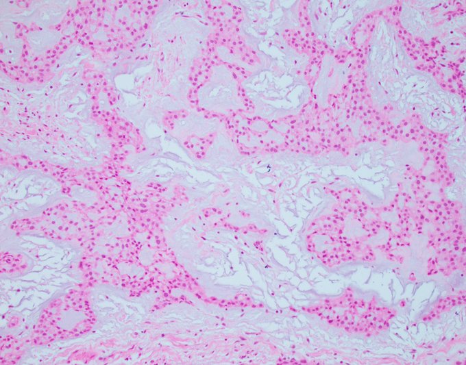
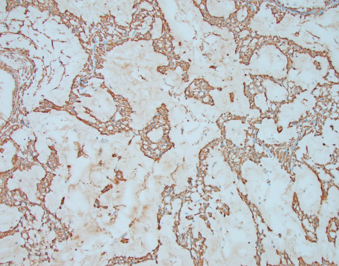
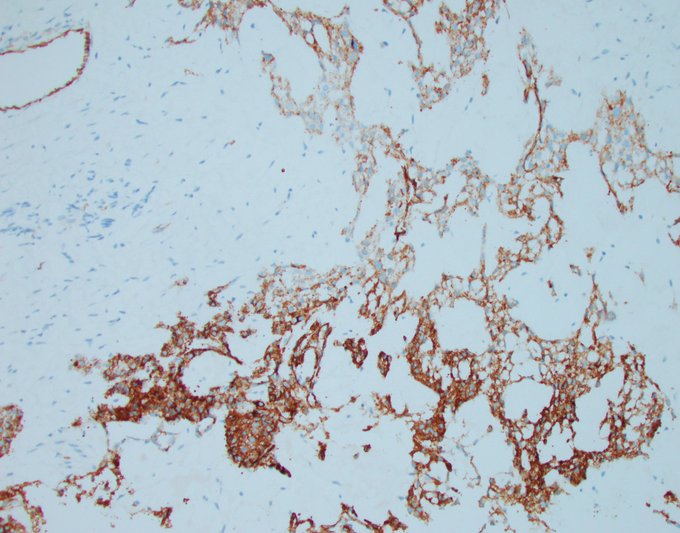
Vascular lesions, mammary or otherwise, can be quite daunting for several reasons. Malignant tumors can look benign, benign tumors can have necrosis and brisk mitotic activity, and heterogeneity is to be expected, which makes diagnosing a vascular neoplasm on core biopsy all the

9
21
71
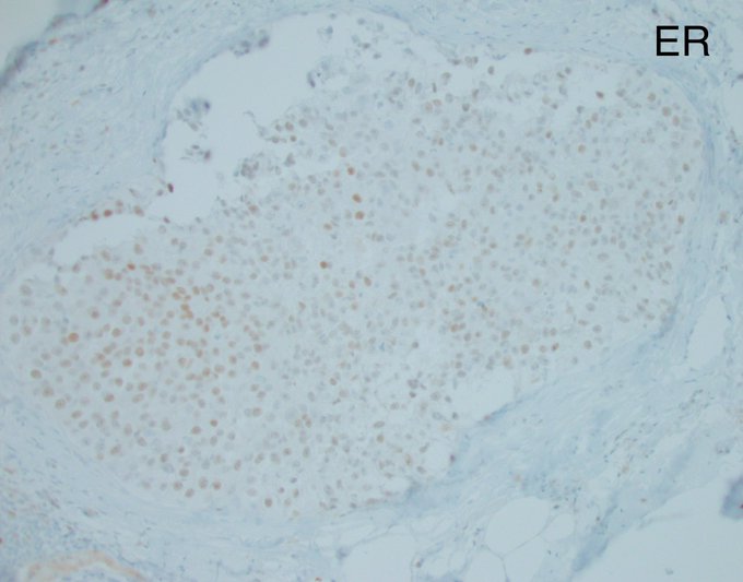
As a follow up to yesterday’s post, CK5/6 and ER are not very useful and can be confusing when the intraductal proliferation consists of a pure population of intermediate and high grade nuclei. DCIS can show basal-like immunophenotype (CK5/6 positive) and can be ER variable to
5
19
71
Does anything grab your attention in this duct with DCIS? if yes, what is it and what do you think is happening?
@washupathedu
@wusm_pathology
#breastpath
#PathTwitter
#PathX
20
17
74

24
19
73
It is wise to remember (and remind clinicians) that morphologic overlap in pathology is a rule that cannot be broken. Keeping an open mind about unconventional patterns of disease and recalling them for future reference is the very essence of expertise.
Behold the gynecomastoid
2
19
73
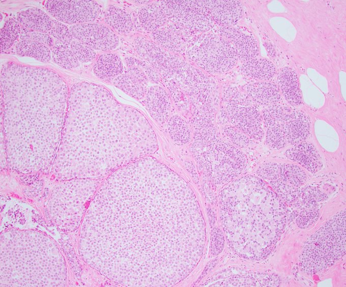
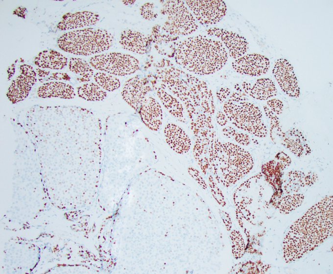
Classic LCIS progressing to Florid LCIS, and losing ER in the process.
@wusm_pathology
@washupathedu
#breastpath
#PathTwitter
#pathology
4
21
72
As many of you have pointed out, this tumor is composed of malignant epithelium and malignant stroma, which is diagnostic of metaplastic carcinoma, and more specifically carcinosarcoma given the collision and lack of smooth transition between the carcinomatous and sarcomatous
8
18
72
Let's open the doors of controversy. My relationship with E-cadherin is strained, to say the least, and I will often rehash my thoughts on this matter to whoever is willing to listen and read. To me, lobular and ductal phenotypes are far from black-and-white varieties with
16
29
73
Thought I was looking at a breast biopsy. Ended up counting glomeruli :)
#PathTwitter
#breastpath
@washupathedu
@wusm_pathology
10
16
71
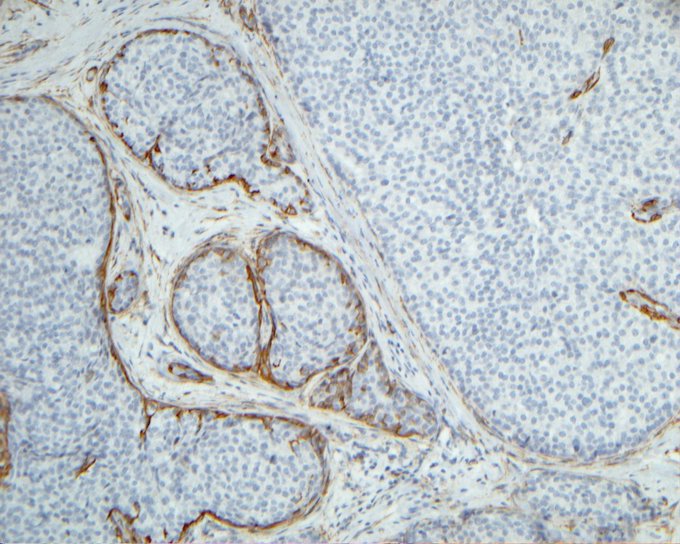
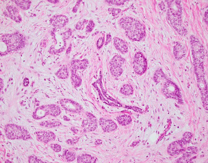
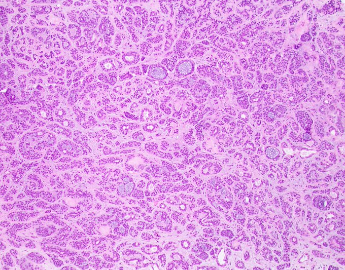
Moving on with another example of tumors with a dual cell population.
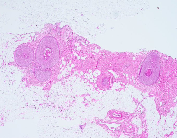
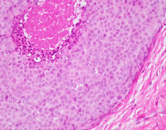
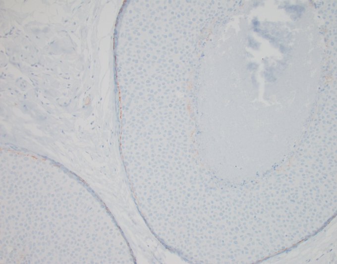
This is the most famous of the bunch, classic adenoid cystic carcinoma.
In contrast to yesterday’s highly infiltrative tumor, this one is, contrary to expectations, well circumscribed, which is what prompted
2
15
71
Fascinating case of an 83-year-old woman with a 6 cm breast mass. What is your diagnosis? comment or enter the poll below
@wusm_pathology
@washupathedu
#PathTwitter
#BreastPath
1
28
70
In my last tweet, two-thirds of the participants correctly diagnosed the focus depicted in the image as non-invasive, despite the lack of myoepithelial cells. The reason is that the focus lies within the previous biopsy site environment (fat necrosis, reactive fibrosis,
4
16
71
I can’t call this a Tweetorial because of the inherent controversy of the subject matter, so I’ll just call it a Saturday opinion piece.
Let’s get granular with lobular. The images I shared a couple of days ago show what to me looks like ALH/LCIS in a duct, and invasive lobular
4
26
71
Back to the basics Tuesday.
This patient has extensive high grade DCIS, but that’s not the question.
The question is: how many ducts and how many lobular units do you see in this low power (2x) snapshot?
Is it all ducts? Is it one big TDLU? is it one duct and multiple
9
15
68
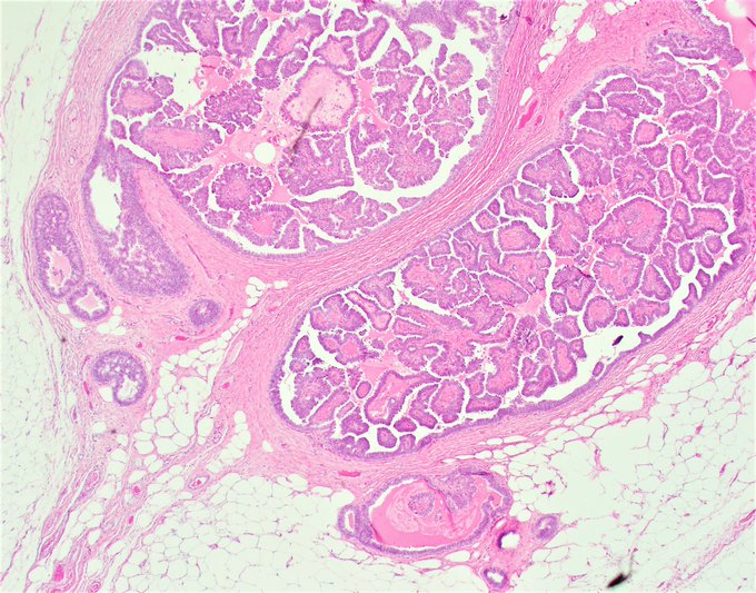
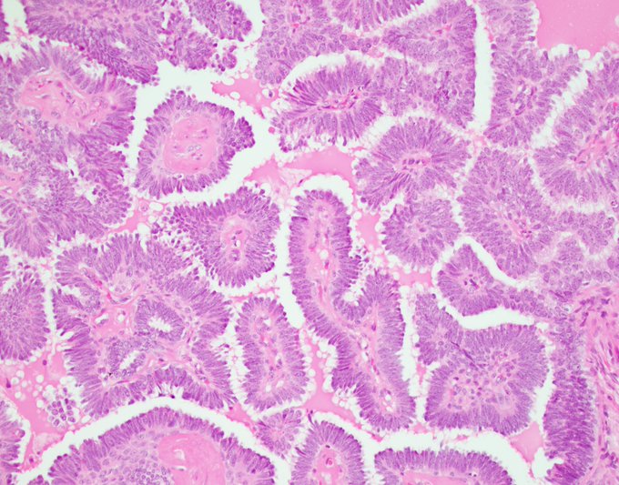
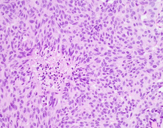
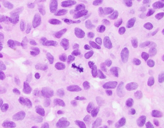
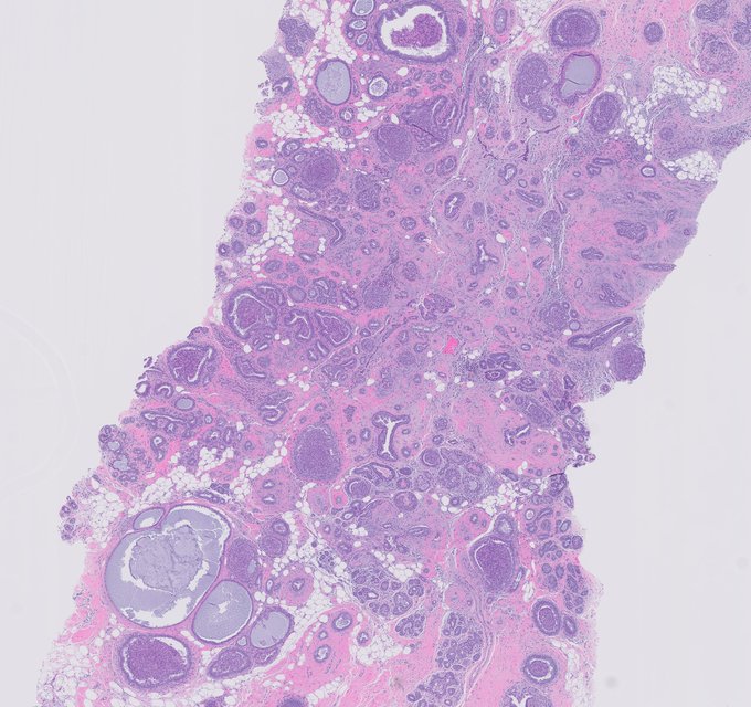
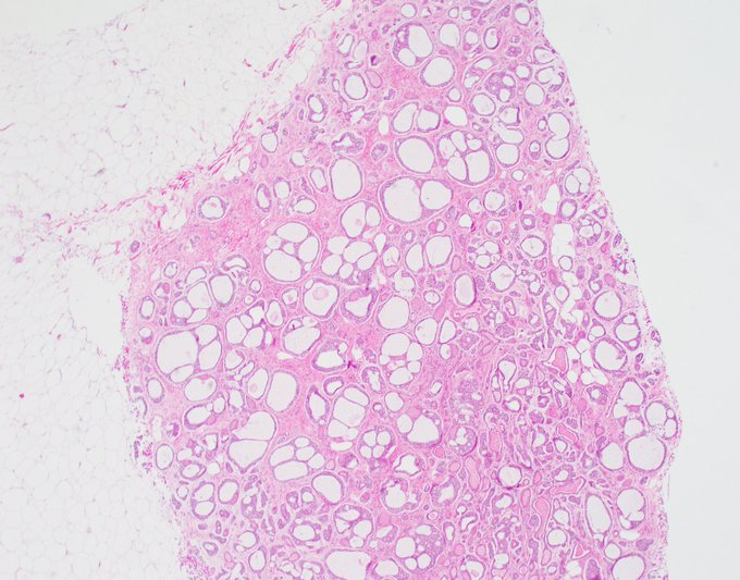
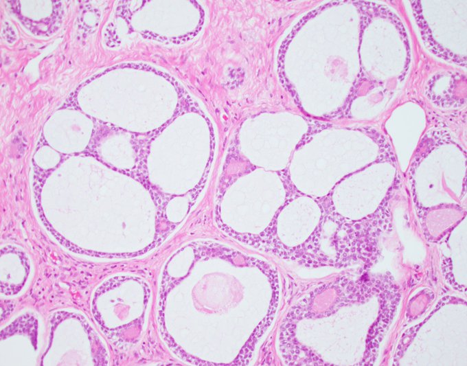
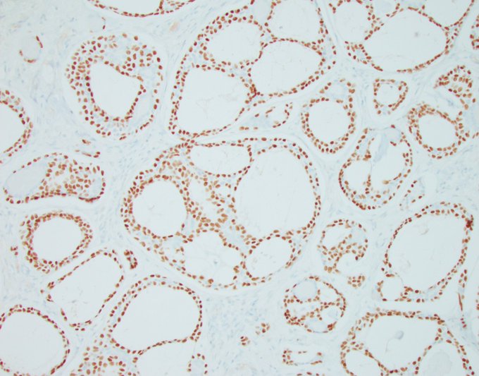
Head-scratcher Tuesday.
This consult case befuddled me for a long time until I did one thing.
Here’s the story it came with. Latin American lady with a retroareolar mass, possibly painful but unsure, slow growing over the past 2-3 years.
Let the guesses and work-ups begin!
17
25
67
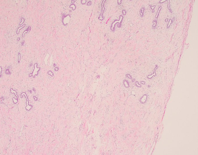
18 year old patient. 8 cm mass.
The pathologist’s question was is this PASH or is this fibroadenoma.
What is your answer?
@wusm_pathology
@washupathedu
#breastpath
#PathTwitter
#PathX

36
19
69
Wrapping up my
#BSTpath
week with this cutie. Not in the breast this time, for a change.
Subungual painful mass, came with a clinical diagnosis of glomus tumor. But glomus tumors have a very characteristic image in my mind with prominent vasculature and sheets of the most
3
14
69
Let’s kick off the new year with everyone’s favorite tumor, the infamous invasive lobular carcinoma masquerading as something it isn’t.
One of the first cases I shared in April of last year was a lobular cancer that looked like fat necrosis. That one was scary, but it doesn’t

9
16
69
Patient in her 30s with at least two lobulated but irregular right breast masses. Negative history.
What do you think?
@wusm_pathology
@washupathedu
#breastpath
#PathTwitter
#PathX
21
21
66
Subtle infiltrative patterns of invasive carcinoma are not always lobular. Sometimes ductal is just as sneaky and treacherous. The key here is to train your eye to recognize on low power slight deviations from normal and mild increases in cellularity, then zoom in and identify.
7
14
66
Two lobular units. What's the difference between them?
#PathTwitter
#breastpath
@washupathedu
@wusm_pathology
12
11
67