
Arkana Laboratories
@arkanalabs
Followers
11K
Following
10K
Media
3K
Statuses
7K
Renal pathology & neuromuscular pathology laboratory focused on teaching others about rare diseases and improving patient care. #renalpath #pathtwitter #pathx
Little Rock, AR
Joined September 2012
Have you integrated APOL1genetic testing into your practice? 🔬✨Discover the No-Cost APOL1 Genotyping Program for eligible patients sponsored by Vertex Pharmaceuticals — helping you deliver precision care without added cost. Learn more today! ⬇️ #GeneticTesting
1
0
8
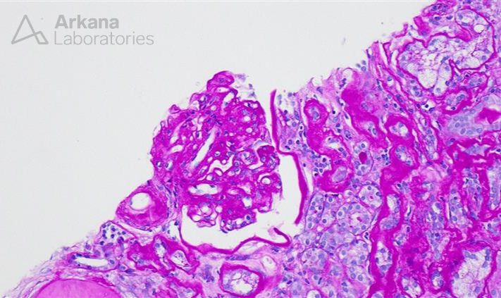
What are the two major changes seen in this image from an allograft kidney and what are these findings concerning for? #DiagnoseThis #pathology #renal #kidneypath

8
8
31
Dr. Stephen Bonsib is back with another episode on ciliopathies, as part of his series Chronicles of Cystic Kidney Disease, which investigates developmental abnormalities and cystic kidney diseases. #renal #kidneypath #pathology https://t.co/YBF6Ra2lQo
0
1
8
Severe acute tubular injury and beaded casts that stain positive for myoglobin in a case of myoglobin cast nephropathy. #renalpath #pathology #kidneypath #pathx #renalx


0
9
28
The renal biopsy adequacy crisis isn't slowing down. For insight on how the renal community might begin to address this challenge, listen to our podcast with @nephrosharma, @Tiff_Caza, @TWhittier_RUSH, @vandyniyyar, and @VelezNephHepato. https://t.co/xw26GmR6oX
0
7
13
ANSWER: A, Autosomal recessive inheritance Nail-Patella syndrome is a rare autosomal dominant disease with an incidence of approximately 1/50,000. The majority of cases are due to a mutation in LMX1B located at chromosome 9q34.1. The gene is important in development as it
0
0
1
ANSWER: A, Autosomal recessive inheritance Nail-Patella syndrome is a rare autosomal dominant disease with an incidence of approximately 1/50,000. The majority of cases are due to a mutation in LMX1B located at chromosome 9q34.1. The gene is important in development as it
All of the following are features of Nail-Patella syndrome except: A: Autosomal recessive B: Mutation in LMX1B C: Banded collagen by EM D: Absent/dystrophic nails #pathX #pathtwitter #renalX #kidneypath
0
1
3
A case of fibrillary glomerulopathy showing smudgy mesangial/capillary loop staining for IgG, positive DNAJB9 staining and fibrils on EM. Superimposed on diabetic nephropathy with KW nodules. #renalpath #pathology #renal #pathx #pathtwitter #kidneypath


0
11
30
All of the following are features of Nail-Patella syndrome except: A: Autosomal recessive B: Mutation in LMX1B C: Banded collagen by EM D: Absent/dystrophic nails #pathX #pathtwitter #renalX #kidneypath
1
0
4
The answer is: amyloidosis. To learn more, head to our blog: https://t.co/6MWAnFQ4kR
#neuropath #neuromuscular #neuronotes #pathology #pathX

arkanalabs.com
Clinical History:The patient has numerous complaints including weakness, myalgia, arthralgia, back pain radiating down bilateral legs, cramps and twitching, imbalance,…
0
0
0
Beautiful hyaline thrombi in a case of cryoglobulinemic glomerulonephritis. This patient has active hepatitis C. #renal #pathology #renalpath #pathx #pathtwitter #kidneypath

0
18
60
A case of AA amyloidosis. This biopsy is from a homeless patient with numerous abscesses of the arms and legs, consistent with infected skin popping wounds due to IV drug use. #pathx #pathtwitter #renalpath #renal


1
12
52
The patient has numerous complaints including weakness, myalgia, arthralgia, back pain radiating down bilateral legs, cramps and twitching, imbalance, speech disorder, difficulty walking, and numbness. His past medical history is remarkable for gout and he is taking both


1
0
2
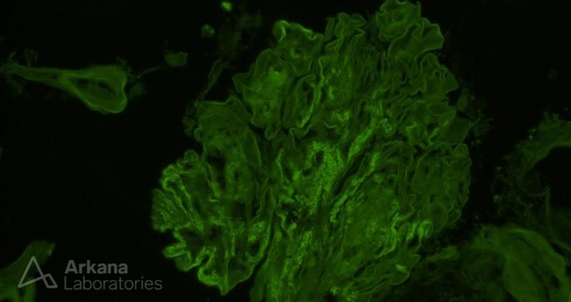
A sample from the pathology teaching slide set on Hepatitis B Infection-Related Glomerulonephritis, jointly prepared by Arkana Laboratories and @goKDIGO. https://t.co/KwXMfV3nTh
#KDIGO #pathology #renal #renalpath

0
10
26
The light microscopic image depicts an artery with a cholesterol embolus. Approximately three-quarters of cases demonstrating atheromatous emboli are iatrogenic in nature and due to invasive, specifically vascular, procedures such as coronary artery bypass, aortic repair (i.e.
0
0
0
The light microscopic image depicts an artery with a cholesterol embolus. Approximately three-quarters of cases demonstrating atheromatous emboli are iatrogenic in nature and due to invasive, specifically vascular, procedures such as coronary artery bypass, aortic repair (i.e.
What is your diagnosis and what are some possible precipitating events that may lead to this finding? #DiagnoseThis #pathology #renal #kidneypath

1
4
12
Here is today’s #eyeSCANdy! Glomerular tuft with overall intact architecture. Discrete masses of specular amyloid interrupt smooth uninvolved segments of capillary loop. Photo courtesy of Dr. Stephen Bonsib. #renal #pathology

0
1
7
Electron microscopy can be helpful in the diagnosis of cryoglobulinemic glomerulonephritis. This renal biopsy (Fig. 1) shows a membranoproliferative pattern glomerulonephritis in which the extent of endocapillary hypercellularity and glomerular capillary double contour formation


0
14
55
Are you aware of and up to date on the growing renal biopsy adequacy crisis? Click the link below to listen to @nephrosharma, @Tiff_Caza, @TWhittier_RUSH, @vandyniyyar, and @VelezNephHepato discuss how we might address this challenge. https://t.co/xw26GmQyzp
0
3
12
What is your diagnosis and what are some possible precipitating events that may lead to this finding? #DiagnoseThis #pathology #renal #kidneypath
7
6
46
ANSWER: B Membranous glomerulonephritis (MGN) is the most commonly described glomerular disease in IgG4-related disease. MGN was present in about 7% of cases of IgG4-related tubulointerstitial nephritis from two biopsy series studies. Reference: Sethi S, et al.
0
0
1