

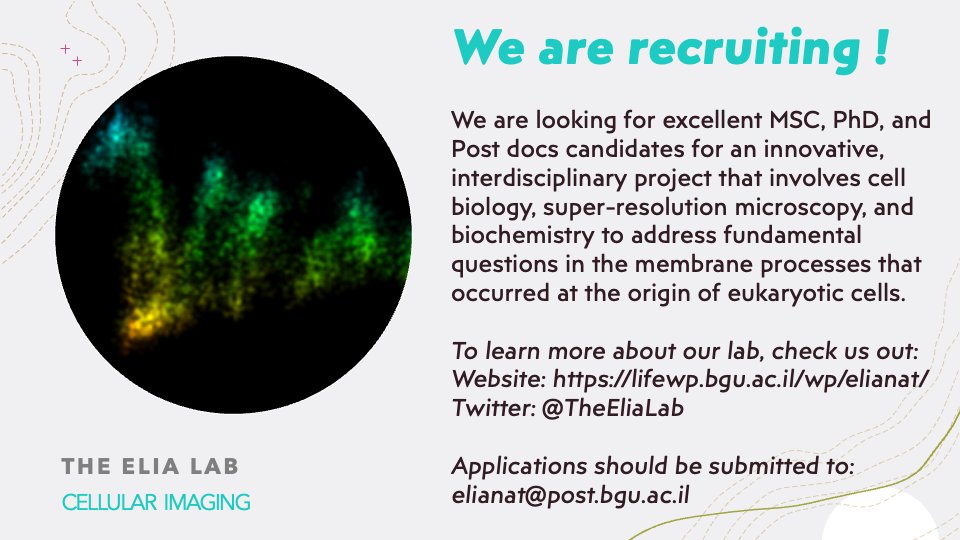
The Elia Lab
@TheEliaLab
Followers
425
Following
794
Statuses
384
Our moto is to advance state-of-the-art microscopy techniques in order to resolve how molecular machines organize in cells to execute their biological function
ישראל
Joined February 2019

RT @EuroBioImaging: Check out this amazing lightsheet #imaging work and catch up on all the news from @DBioimaging below⤵️
0
4
0
RT @PIHanson: Now out at PNAS - excited to share our work on how ESCRTs protect lysosomes from stress-induced membrane rupture, enhancing l…
0
30
0
RT @labs_mann: Excited to share our study on BioRxiv! We developed a spatial proteomics approach combining multiplexed imaging & deep visua…
0
58
0
RT @Dey_Gautam: Science is bloody hard but filled with unexpected joys - the joy of watching colleagues @dudin_o @SchwabYannick turn into l…
0
20
0
RT @PLOSBiology: AAA-ATPase VPS4 helps separate daughter cells at the end of #cytokinesis, but why 2 paralogs? @TheEliaLab shows that #VPS4…
0
2
0
RT @EuroBioImaging: At this week's #VirtualPub, learn about "Tools from AI4Life that anyone can use" with speakers Anna Kreshuk @ilastik_te…
0
10
0
RT @TechnionLive: בתקופת השיקום במרכז הרפואי לשיקום לוינשטיין קיבלה נטע סד ייחודי, שתוכנן ויוצר במיוחד עבורה בטכניון במעבדתה של @DanaSolav…
0
3
0
1
0
1
RT @aaandmoore: It's not a deep sea dweller. This is actually the endoplasmic reticulum of a cell, segmented and tracked through time. htt…
0
145
0

@LabMizrahi @NatureMicrobiol @GurLab @JensWalter15 It was a pleasure, as usual, to contribute our five cents to this elegant work
1
0
1
RT @LabMizrahi: 📚🌐Thrilled to share our newly published paper in @NatureMicrobiol in which we unravel how plasmids can tip the scales in mi…
0
106
0
RT @AQLMMBL: The North Atlantic Microscopy Society (NAMS)@NAMS_Microscopy is sponsoring two $500 grants for minority students to attend AQL…
0
11
0
RT @ProjectNemi: 🦠Thylakoid membranes with #phycobilisomes of #cyanobacteria imaged by Dr. Laura van Bezouwen, Utrecht University (@UniUtre…
0
12
0