Arizona Ultrasound
@SonoStache
Followers
3K
Following
1K
Statuses
495
This is the official twitter feed for all things Arizona and Emergency Ultrasound! Program Director: Dr. Srikar Adhikari
Tucson, AZ
Joined June 2014
0
0
2
0
4
10
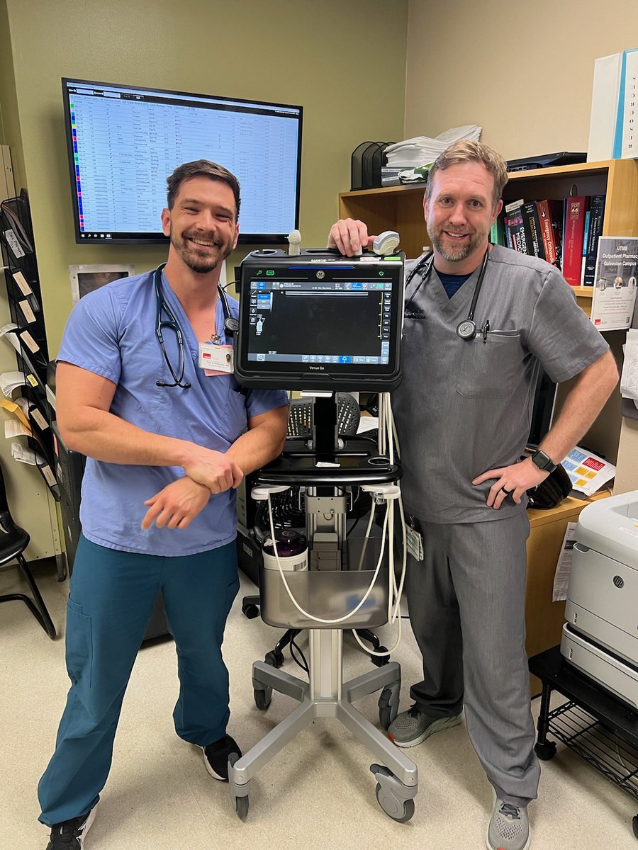
Want to learn #POCUS or hang out with us for a month? We love visiting rotators. Come meet the team!

0
1
3

What do you think is happening in this ultrasound? Check out for the answer!
3
3
13
Time for another case of the week! Lets see who knows what this abnormality is... Be sure and double check yourself at
1
0
1
What abnormality is seen on this young, pregnant female? Learn more at:
0
1
0

Adhikari's Image of the Week: G-tube placement confirmation. Once visualized in the stomach, color Doppler can be applied over the tube to enhance visualization by motion artifact during gentle tube oscillation. #POCUS
0
3
8
Join us today! “Ultrasound in EMS 101 For Medical Directors - From Idea to Successful Implementation” #NAEMSP2023 @NAEMSP
0
0
4
Adhikari's Image of the Wk: Adenomyomatosis. Can be focal or diffuse. US shows wall thickening w/multiple small cystic pockets (Rokitansky-Aschoff sinuses) and comet-tail artifacts representing cholesterol aggregates. #POCUS
1
17
49
RT @SAEMAEUS: Today's #SonoGames22 Round 0 Infographic comes from @scan_yes and reviews an important paper by researchers from @UArizonaEM,…
0
5
0
Adhikari's Image of the Week: US features are non-specific: Echogenic medullary pyramids, some may cast posterior shadowing. Predominantly bilateral. Distinguishing between medullary nephrocalcinosis can be difficult, as many times these conditions coexist. #POCUS
0
4
5
RT @ResaELewiss: Development and Validation of a Point-of-Care-Ultrasound Image Quality Assessment Tool: The POCUS IQ Scale
0
23
0

Adhikari’s Image of the week: Malignant thyroid nodule. Hypoechoic relative to adjacent thyroid tissue. A nodule that is ‘taller than wide’ in shape, increases risk of malignancy. Can also have irregular edges. Tend to demonstrate intranodular vascularity. #POCUS
0
8
11