

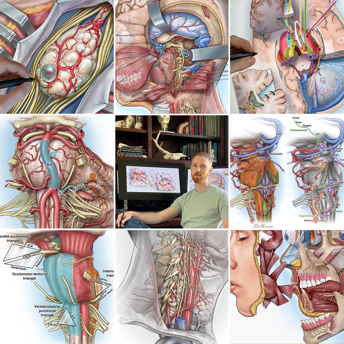
Peter M. Lawrence
@PeterMLawrence1
Followers
41K
Following
20K
Statuses
1K
🧠✍🏻 Certified medical illustrator @barrowneuro
Phoenix, AZ
Joined August 2013
Another year of doing what I love. Thanks for the support, everyone 🧠✍🏻 #neuroscience #illustration
203
1K
13K
RT @PeterMLawrence1: Made a few changes to some illustrations depicting the endoscopic endonasal eustachian tube anterolateral mobilization…
0
26
0
RT @BarrowNeuro: In Nov., we hosted our first funded #neurology observer at Barrow, thanks to the Franke #GlobalNeuroscience Education Cent…
0
1
0
This drawing was a lot of fun to make 🍇✍️

Watch the Seven Series video showing this right temporal-parietal craniotomy, transsulcal approach, resection of giant periventricular cavernous malformation, on our website @BarrowNeuro by clicking on this link…

2
2
54
RT @mtlawton: This collage reminds me of standing in the Sistine Chapel and looking up. Magnificent illustrations, and thank you @PeterMLaw…
0
8
0
1
0
1
0
0
1
These BSCM approach overview illustrations are always a challenge to illustrate, but some of my favorite from the Seven Cavernomas series. Gotta love the "glass" arrows for the approach trajectories #Neuroscience #MedTwitter
Watch the Seven Series video showing this extended retrosigmoid craniotomy, trans-middle cerebellar peduncle approach, resection of recurrent pontine cavernous malformation, on our website @BarrowNeuro by clicking on this link…

0
12
88
Always neat to illustrate an AVM 🧠✍🏻


2
16
203
One of the most beautiful anatomical treatises ever published, Bourgery & Jacob’s Atlas of Human Anatomy and Surgery #neuro #medicalillustration

From “The Complete treatise of human anatomy” by J.M. Bourgery and N. H. Jacob, published in Paris between 1831 and 1854. The work is monumental: eight volumes of text totalling 2108 pages, and atlas volumes with 725 plates, representing a total of 3750 figures. @PeterMLawrence1


2
22
183
RT @mtlawton: #SevenCavernomas is down the home stretch. I think this was the last of the illustrations to complete the art program. The ma…
0
1
0

0
64
0
The posterior cerebral artery (PCA) has five segments: P1, pre-communicating segment, originates at the end of the basilar artery & terminates at the posterior communicating artery (green) P2, post-communicating segment, begins after the posterior communicating artery (PCOM) and loops around the midbrain & terminates at the origin of the lateral occipital artery as it enters the quadrigeminal cistern (blue) P3, quadrigeminal segment, which runs through the quadrigeminal cistern in a posteromedial direction (purple) P4, cortical segment, located within the sulci of the occipital lobe (yellow) P5, terminal branches (orange) #neuroeducacion #MedTwitter @OGdukeneurosurg @Innov_Medicine
1
30
149
RT @mtlawton: Watch the Seven Series video showing this frontotemporal craniotomy, transsylvian-transinsular approach, resection of giant l…
0
11
0
1
35
232
WIP, superior view of skull base depicting both posterior cerebral artery (PCA) and superior cerebellar artery (SCA) segments, mesencephalon, and other relevant anatomy #MedTwitter #Neuroscience #wacom

4
37
242
This was one of my favorite illustrations I created this year 🧠✍️
Watch the Seven Series video showing this extended retrosigmoid craniotomy, trans-middle cerebellar peduncle approach, resection of petrosal cerebellar and middle peduncular pontine cavernous malformations, on our website @BarrowNeuro by clicking:

0
17
185
The posterior inferior cerebellar artery (PICA) has five segments: P1, anterior medullary: located in front of the medulla (blue) P2, lateral medullary: runs alongside the medulla and ends at the origin of the glossopharyngeal, vagal, and accessory nerves (purple) P3, tonsillomedullary: runs around the caudal half of the cerebellar tonsil (green) P4, telovelotonsillar: runs in the cleft between the tela choroidea and the inferior medullary velum, and the superior pole of the cerebellar tonsil (orange) P5, cortical: distributed to the cerebellar surface (yellow) Illustrated for @mtlawton ‘s 7 Cavernomas #Neuroscience #MedTwitter #NeuroTwitter @OGdukeneurosurg @Innov_Medicine
1
131
538