
Dr. OMID BANDARCHI
@OBandarchi
Followers
37K
Following
10K
Statuses
4K
●M.D. RADIOLOGIST ●PERSONALIZED MEDICINE EXPERT ●MEMBER 𝓸𝓯 IDRA ●امید بندرچی
𝐈𝐑𝐀𝐍
Joined January 2022
🛑MCL bursa located along the middle 1/3 of the medial aspect of knee between superficial & deep portions of MCL. 🛑MCL bursitis is rare. Calcification of MCL is also rare but found either inside the bursa (calcific bursitis) or outside of it confused with meniscal tears,gout,..

0
19
132
Anastomoses around knee. POPLITEAL A. bifurcates into ANT & POST TIBIAL ARTERIES. 🛑There're 4 GENICULATE BRANCHES, a SUP & INF both MEDIALLY & LATERALLY. Also 2 supplementary arteries: DESCENDING GENICULATE A.(a branch of femoral a). ANTERIOR RECURRENT branch of Ant.Tibial a

0
21
88
🛑American Spinal Injury Association(ASIA)impairment scale describes a person's functional impairment as a result of Spinal Cord Injury (SCI) & indicates how much sensation a person feels after light touch & a pin prick at multiple points on body & tests key motions on both sides

0
47
131
🛑Three arches of the foot shown. Furthermore, this image beautifully shows that the 'TRANSVERSE ARCH" of the foot receives & transfers loads during gait. Transverse arch is marked in red and it is contact with the ground during terminal stance.

0
39
180
🛑Distal radius fractures include a group of fractures such as Colles Fx, Smith Fx, Barton Fx, Lister's tubercle Fx, chauffeur Fx. Radiological measurements are useful for diagnosis and follow-up of the treatment. Images : Source:

0
25
148
3 types of HIP DISLOCATION: Central, Posterior, Anterior. The key to differentiate between them is the femoral position. For example in posterior type which is the most common, femur is adducted & fixed in internal rotation. Anterior dislocation has 2 types👉more in the image

0
47
182
🛑Columns vs Walls of acetabulum : The anterior & posterior WALLS of the acetabulum extend from corresponding COLUMNS ( anterior column & posterior column), consequently forming acetabular cup, and stabilize the hip joint. The posterior wall is larger and extends more laterally.

0
55
250
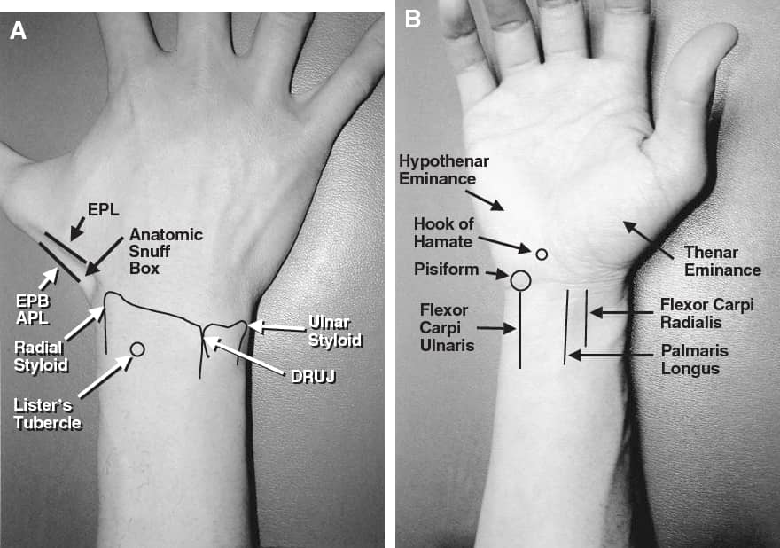
🛑 SURFACE ANATOMY OF THE WRIST ! The wrist is a complex, elegant joint that serves as a key linkage between the forearm & the hand. A basic understanding of wrist and related anatomy allows for appreciation of the biomechanics of wrist movement
0
93
411
Posterior ankle impingement usually develops in jumpers, ballet dancer,.etc secondary to repeated/forced ankle plantar flexion,leads to soft tissue compression. Lateral talar process (If unusually large, called Steida processor) or an os trigonum can also cause post. impingement

0
26
160
🛑Majority of acute ELBOW conditions in emergency setting can be detected on plain xray. 🛑Xray series includes: ✅AP ✅LATERAL ✅OBLIQUE RADIALHEAD- CAPITELLAR The last one help detecting subtle Fx(s) by removing osseous overlap of radial head & coronoid

1
76
501
LIGAMENTS OF STERNOCLAVICULAR & ACROMIOCLAVICULAR JOINTS beautifully shown👍 As well as 2 CLASSIFICATIONS (Tossy and Rockwood ) OF INJURIES TO ACROMIOCLAVICULAR LIGAMENTS mentioned👌

0
28
160
ILIOINGUINAL NERVE provide sensory innervation to the skin of the upper anteromedial thigh & partially the external genitalia. Moreover, it provides motor innervation to internal abdominal oblique & transversus abdominis muscles. Also genital branch of the genitofemoral nerve..

0
10
36
🟥 MALUNION , DELAYED UNION , & NONUNION briefly explained . These entities considered as complications of fractures . Furthermore, the different types of NONUNION shown and the importance of correct diagnosis of these complications mentioned.

0
74
300
🛑All about the knee joint: structure, kinematics,..etc. From: Netter’s concise orthopaedic anatomy

0
60
320
Scapholunate dissociation (SLD) is a symptomatic wrist dysfunction that results from partial or total rupture of scapholunate ligamentous complex which consists of 3 structures: 2 scapholunate lig(s)[palmar & dorsal] & proximal fibrocartilaginous membrane

0
29
148
A few radiological features of bones of foot illustrated. Peroneus brevis tendon attached to styloid process of the base of 5th metatarsal. The oblique epiphyseal line here should NOT be confused with a Fx of styloid process, which is usually transverse, termed a Jones fractue.

0
33
180
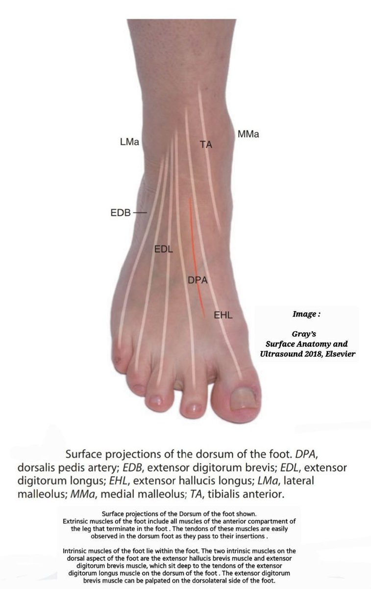
TENDONS SURFACE ANATOMY: DORSUM OF FOOT Tendons of EXTRINSIC muscles of foot observed in the dorsum foot. The two INTRINSIC muscles on dorsal aspect of foot are extensor hallucis brevis & extensor digitorum brevis. The latter can be palpated on the dorsolateral side of the foot.
0
51
243
🛑HIP INVOLVEMENT in OA (Osteoarthritis) vs RA (Rheumatoid arthritis) Hip OA tends to be bilateral & in addition to typical features of OA, superolateral cartilage space narrowing occurs. In RA concentric acetabular cartilage loss, leads to the axial migration of femoral head.

0
33
163
🛑 Comment for tweet of ANB: The size of each ANB calculated by taking its highest axial measurement (highest reading recorded when drawing a straight line from the bone edges) in a standard AP X-ray view.
0
0
4