
Elizabeth Montgomery, MD
@LizMontgomeryMD
Followers
21K
Following
12
Statuses
1K
Gastrointestinal & soft tissue pathologist. Professor of Pathology at University of Miami. Founder of @science_press
Miami FL
Joined July 2018
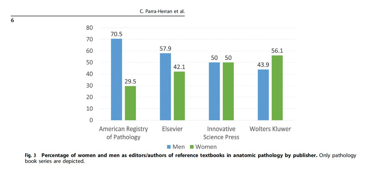
From - Parra-Herran C, Khani F, Wobker SE. Gender distribution of editors and authors of reference textbooks in anatomic pathology. Mod Pathol. 2022 Sep 7. Epub ahead of print. PMID: 36071098. It’s nice that we have gender parity amongst our authors at Innovative Science Press.
5
12
59
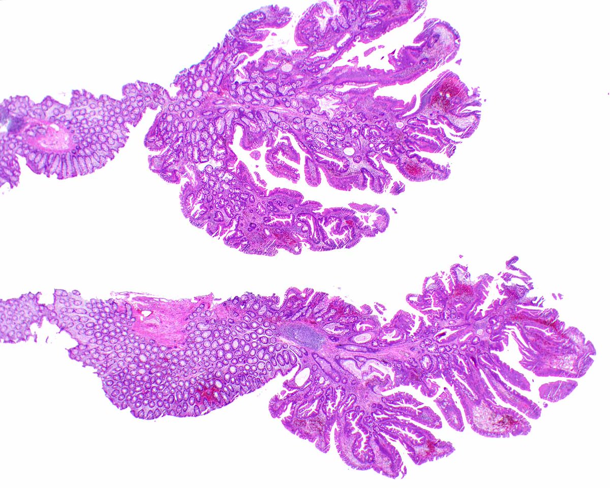
This subtle gastric carcinoma invades the lamina propria (T1a, early gastric adenocarcinoma). The lowly PAS/AB can be helpful in confirming the diagnosis - the tinctorial quality of the mucin is not that of normal or reactive glands. @UMiamiPath @modernpathology @Science_press


1
20
70
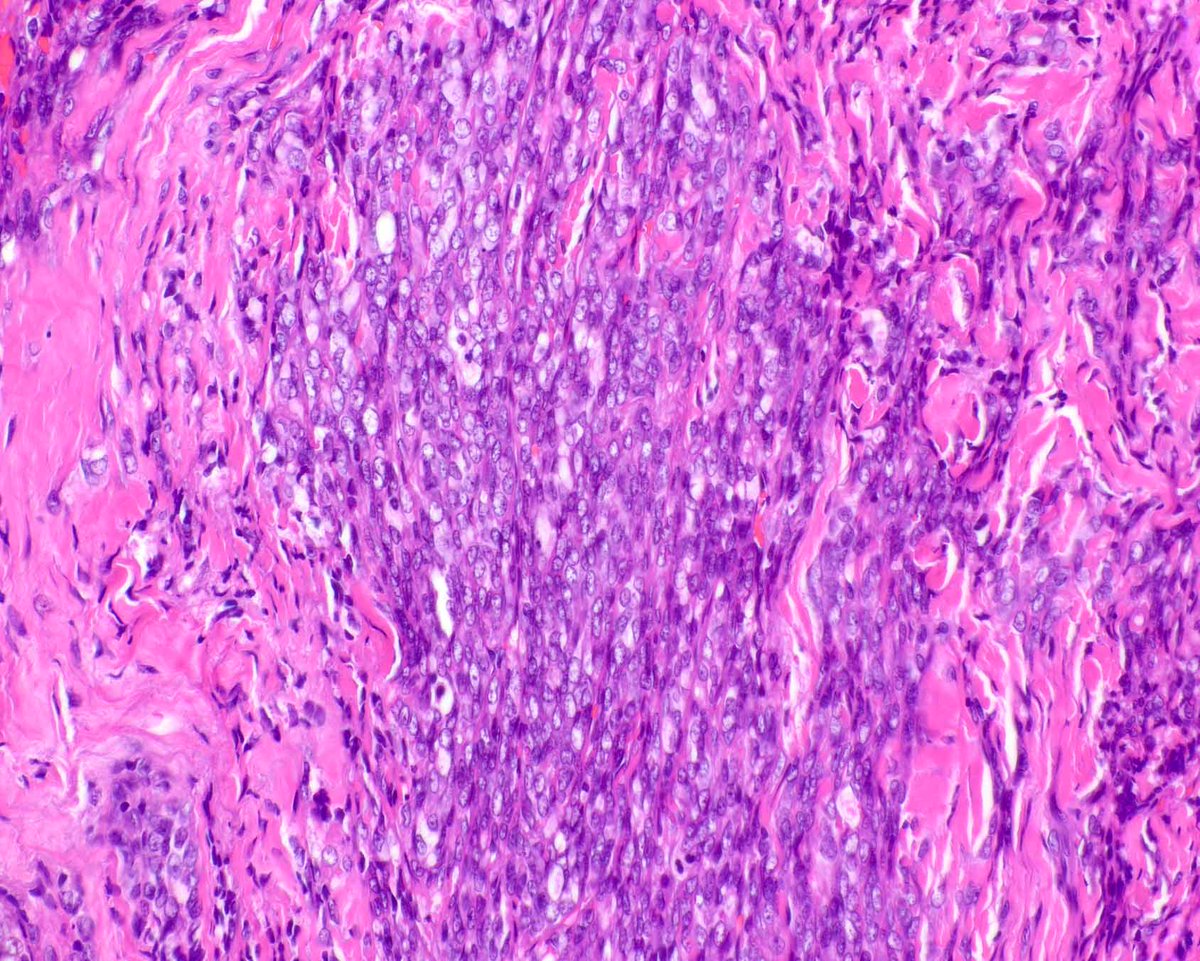
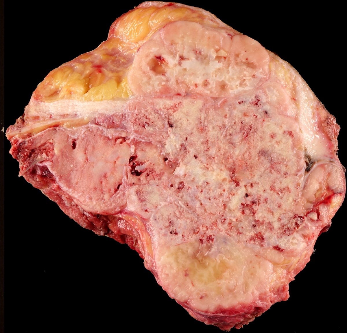
This awful-looking malignant neoplasm proved to be a malignant solitary fibrous tumor on molecular studies - I was not the one clever enough to order the STAT6, but it is patchy - the detection of the NAB2::STAT6 fusion was important.

0
0
2
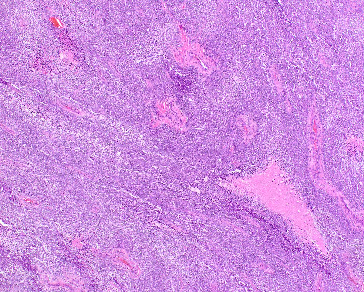
This awful-looking malignant neoplasm proved to be a malignant solitary fibrous tumor on molecular studies - I was not the one clever enough to order the STAT6, but it is patchy - the detection of the NAB2::STAT6 fusion was important.
0
0
5
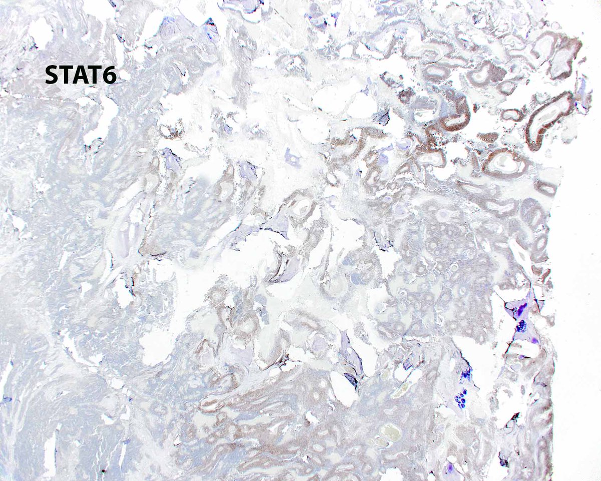
This awful-looking malignant neoplasm proved to be a malignant solitary fibrous tumor on molecular studies - I was not the one clever enough to order the STAT6, but it is patchy - the detection of the NAB2::STAT6 fusion was important.
1
20
68
Please join us for a virtual GI course! Register at

1
4
17
Please join us for a virtual GI course! Register at
0
10
19
Malignant glomus tumor of the small intestine. The collagen type 4 invests each cell.


0
1
8
We are offering a virtual weekend GI and Liver course! Please join us.
0
13
46
Malignant glomus tumor of the small intestine. The collagen type 4 invests each cell.
1
41
156

This mesenchymal chondrosarcoma can be diagnosed on H&E because it displays the round cell component and mature-appearing cartilage, but it can be tough on needle biopsies and NKX2.2 is often reactive! Mod Pathol. 2016 Apr;29(4):370-80. PMID: 26847175. @ModernPathology


2
27
105

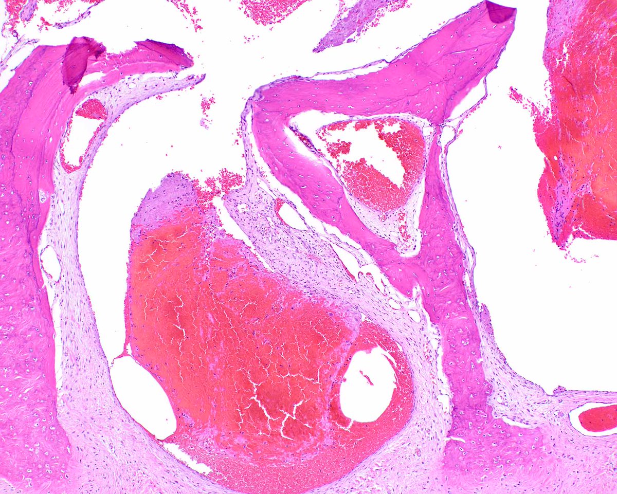
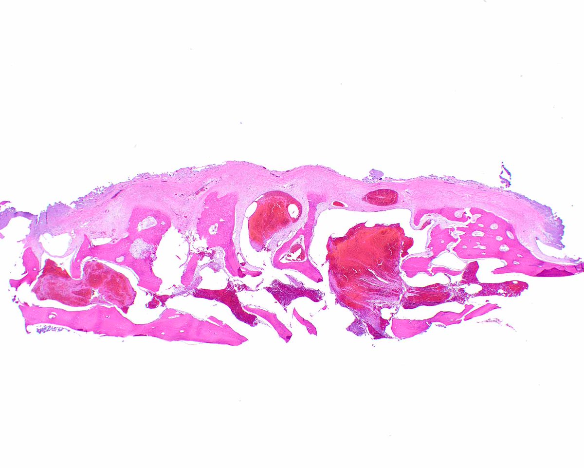
Hemangiomas can give a person a thick skull, although sometimes some of us have one anyway! #UMiamiPath
0
21
89
An incredible esophageal adenosquamous carcinoma arising in Barrett eosphagus with high-grade dysplasia. #UMiamiPath


2
36
126
Spirit off to London 29 April to 2 May for a wonderful gastrointestinal/liver pathology course Also check out this video!
0
1
8
This spectacular succinate dehydrogenase (SDH) deficient gastrointestinal stromal tumor spread to the ovary, where is retains a plexiform pattern. #UMiamiPath


2
37
115
We have an unexpected surgical pathology fellowship opening at The University of Miami for July 2025. Our cases are spectacular (far better than this CMV gastritis) and we have a deep faculty bench. Contact @RhondaYantiss if you are interested at Rky6@med.miami.edu #UMiamiPath
1
26
116
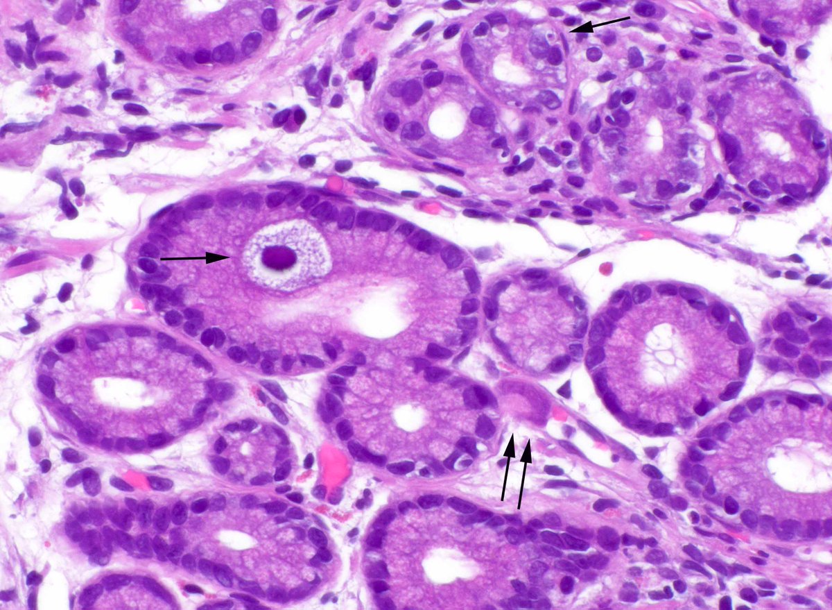
After neoadjuvant chemoradiation even esophageal submucosal glands (arrow) appear atypical. The architecture saves the day. Compare the organized tubules of the benign glands to the disordered treated malignant ones. #UMiamiPath


0
15
97