

EchoTech
@EchoTech_4
Followers
4K
Following
3K
Statuses
3K
"Every image tells a story of love, hope, and new beginnings. #EchoTech"
Yemen
Joined June 2021


44 years old female presented with slow growing painful palpable masses since 1 year at the left lower abdomen and groin. #Echo_Tech


7
9
51
@DrKintaro If just hypertension you will see low vlocity <10cm and venous like waveform will be noted associated with respiratory phasicity , but here there is abnormal pulsatile arterial like waveform which indicate cardiac reflections
Agree 👍
0
2
2
Hypertrophied column of bertin(normal anatomical variant), may mimicking Neoplasm or duplex kidney. US Features of it : - isoechoic to the renal cortex because its extension of it. -normal vascularity (no angiogenesis) -between the two pyramid. #Echo_Tech


0
22
102
Exactly 🤝

@EchoTech_4 As associated with right pleural effusion that is obviously noted in the same sonogram, so it sounds to be reflection of heart disease. Further cardiologist consultation is highly recommended
0
0
0

@Chickoo275 Ok We suspect arteriovenous fistula if we see a typical arterial waveform incthe vein , but this seems to be just reflection for other condition.
1
0
2
Occluded left ICA likely of embolic etiology (look for the pulsatile sign). Note this🪧 if you see a healthy intima media with no signs of atherosclerosis and the vessele is occluded so its mostly embolic as well as you may see a flow in the periphery. #Echo_Tech
49 years female presented with signs of right hemiplegia as Poor balance , weakness , Difficulty walking and speech associated problems. Carotid exam was requested by the clinician , what is your opinion? Note 📝: the vertebral system was unremarkable. #Echo_Tech


0
11
43

1
0
1
Reported as that and highly recommended for followup.

@EchoTech_4 Reactive for follow up after proper treatment.
1
0
6
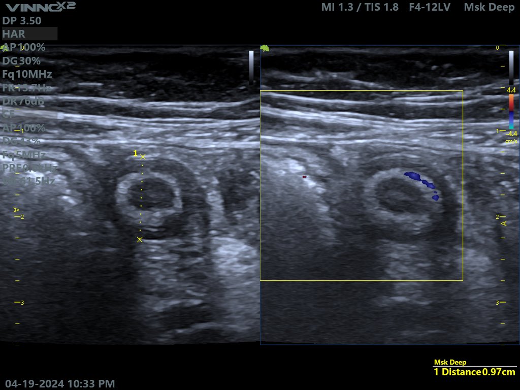
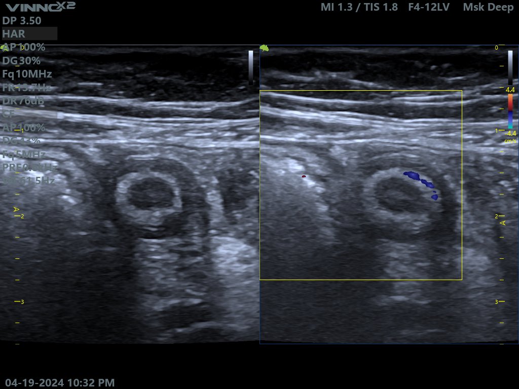
4 years male patient presented by diffuse neck swelling no redness , no pain, no fever at the time of exam. Ultrasound revealed innumerable enlarged cervical lymphnodes in both sides more in the submandibular region This is the largest lymphnode. Your opinion? #Echo_Tech


13
12
62
@lily21392 I think ultrasound is good for this diagnosis and the clinican will be able to get the right diagnosis when he correlate the ultrasound findings with lab and clinical exam.
1
0
1